
1 Physical and Theoretical Chemistry - NanoBioScience, TU Braunschweig, Hans-Sommer-Strasse 10, 38106 Braunschweig, Germany.
g.acuna@tu-bs.de
We report on the development of a bottom-up nanoantenna to enhance the fluorescence intensity in a reduced hot-spot, ready for biological applications. We use self-assembled DNA origami structures as a breadboard where different gold nanoparticle systems consisting of dimers and monomers are positioned with nanometer precision. The dependence of the fluorescence enhancement on nanoparticle size is studied and compared to numerical simulations. A maximum of 100fold intensity enhancement is obtained using 100 nm gold nanoparticles at a gap of 23 nm between the dimer. Additionally, the binding and unbinding of short DNA strands on the hotspot of the nanoantenna is realized, showing the compatibility of this technique with biomolecular assays. The combination of metallic nanoparticles with DNA origami structures with docking points for biological assays paves the way for the development of bottom up inexpensive enhancement chambers for single molecule measurements at high concentrations where processes like DNA sequencing occurs.
Funding by a starting grant (SiMBA) of the European Research Council, the Volkswagen Foundation, and the Center for NanoScience is gratefully acknowledged.
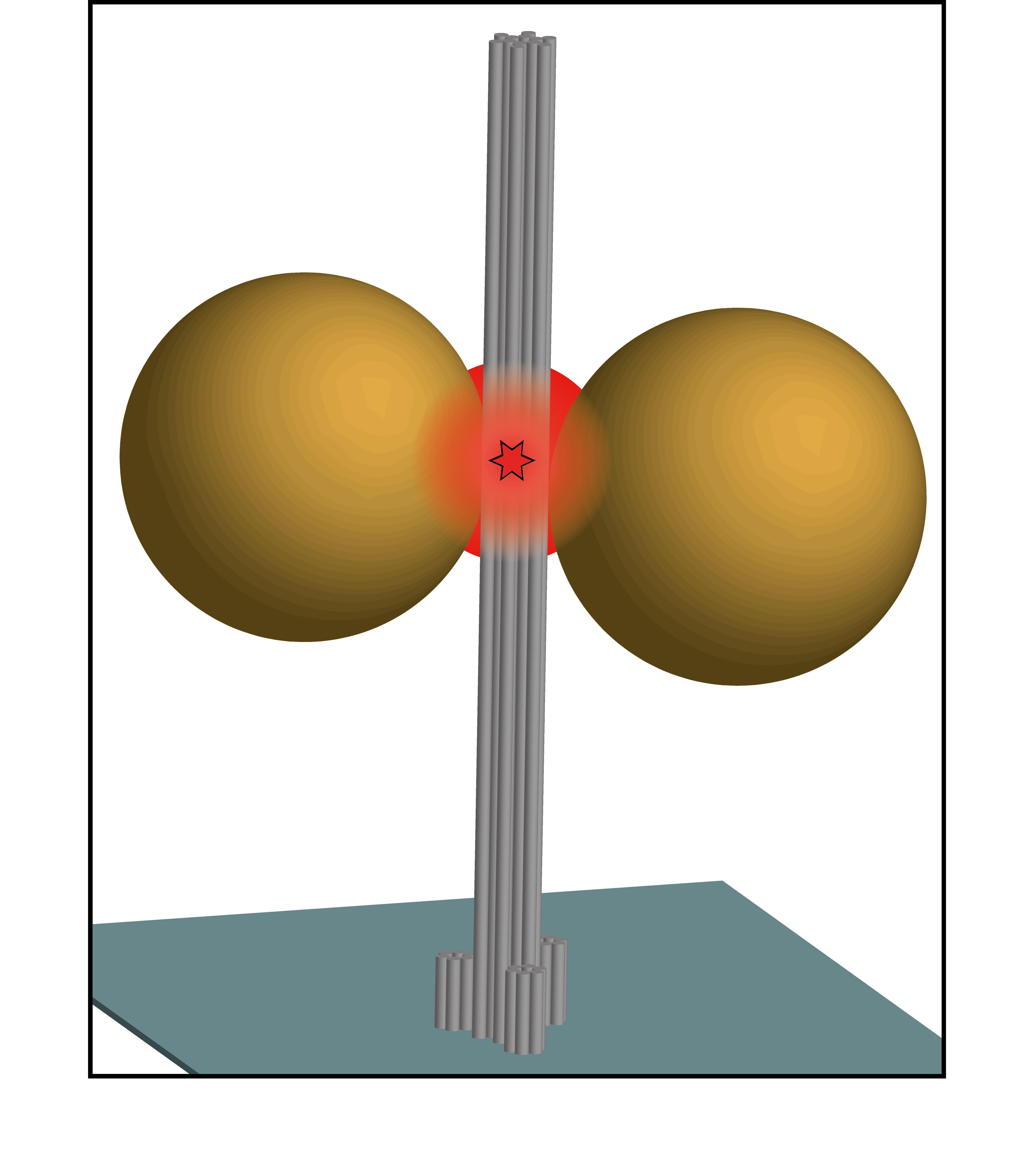
- Fig. 1: Sketch of a dimer nanoantenna based on a DNA origami pillar structure
[1] G. P. Acuna et al., Fluorescence enhancement at docking sites of DNA-directed self-assembled nanoantennas. Science 338, 506-510 (2012).
The Blackett Laboratory, Department of Physics, Imperial College London, London SW7 2AZ UK
* Department of Electrical and Computer Engineering, National University of Singapore, Singapore 117576
** Acad Sci Czech Republic, Int Photon & Elec, Chaberska 57, CR-18251 Prague, Czech Republic
Metallic nanodevices based on surface plasmon polaritons provide new routes to enhance light matter interactions in subwavelength volumes [1], with major applications in molecular sensing [2], light-emitting devices [3] and photovoltaics [4]. In this context, special attention was recently devoted to optical antennas, counterparts of radio and microwave antennas in the optical regime [5]. By reversibly converting propagating electromagnetic waves into localized energy and modifying the emission properties of individual quantum emitters, optical antennas appear to be essential devices to enhance fluorescence spectroscopy [6], Raman spectroscopy [7] and infrared absorption spectroscopy [8]. However, because of their dipolar properties, such optical antennas exhibit a narrow-band response, and hence are not suitable for applications related to multispectral sensing of biomolecules, nonlinear vibrational sensing and nonlinear plasmonics. Despite significant progress, developing robust optical nanodevices with a significant bandwidth of operation remains an open challenge in plasmonics.
In this presentation, we will report a significant step towards tackling this challenge via the use of optical antennas operating in a broad range of frequencies [9]. Our work is based on a log-periodic optical antenna providing high electromagnetic intensity enhancements on a bandwidth of several octaves. Experimental demonstration of surface-enhanced detection using fluorescence, Raman and infrared absorption processes have been recently highlighted, thus opening new opportunities for the development of biosensors enabling multispectral sensing on the same substrate.
[1] S. A. Maier et al, Plasmonics: Fundamentals and Applications; Springer: New York, 2007.
[2] Y. Fu et al, Laser Photonics Rev. 2009, 3, 221.
[3] J. A. Schuller et al, Nat. Mater. 2010, 9, 193.
[4] H. A. Atwater et al, Nat. Mater. 2010, 9, 205.
[5] L. Novotny et al, Nat. Photon. 2011, 5, 83.
[6] A. Kinkhabwala et al, Nat. Photon. 2009, 3, 654.
[7] B. Yan et al, ACS Nano 2009, 3, 1190.
[8] F. Neubrech et al, Phys. Rev. Lett. 2008, 101, 157403.
[9] M. Navarro-Cia et al, ACS Nano 2012, 6, 3537.
[10] H. Aouani et al, ACS Nano 2013, 7, 669.
[11] H. Aouani et al, to be submitted.
Nanoscience Center, University of Jyväskylä, P.O. Box 35, 40014, Jyväskylä, Finland
We experimentally demonstrate the strong coupling between surface plasmon polaritons and the S2 state of β-carotene [1]. The SPPs are excited by prism coupling technique on a thin silver film with β-carotene embedded in a polymer layer on top of that. Rabi splittings with energies 80 and 130 meV are observed in the recorded dispersion relations. Both coupled oscillator model and transfer matrix method are used to fit the experimental results. The scattered radiation of the propagating strongly coupled SPP-S2 hybrids is collected and an increase of the low energy splitting to 120 meV is observed compared to the reflectivity data. In addition, we perform molecule excitation by laser, and analyzed the emission patterns revealing clear surface plasmon coupled fluorescence of β-carotene. By increasing the concentration of β-carotene we are able to collect also surface plasmon coupled Raman scattering. This study substantially extends the SPP-molecular excitation strong coupling studies to biomolecules, and energy transfer and coupling properties of excited states of carotenoids.
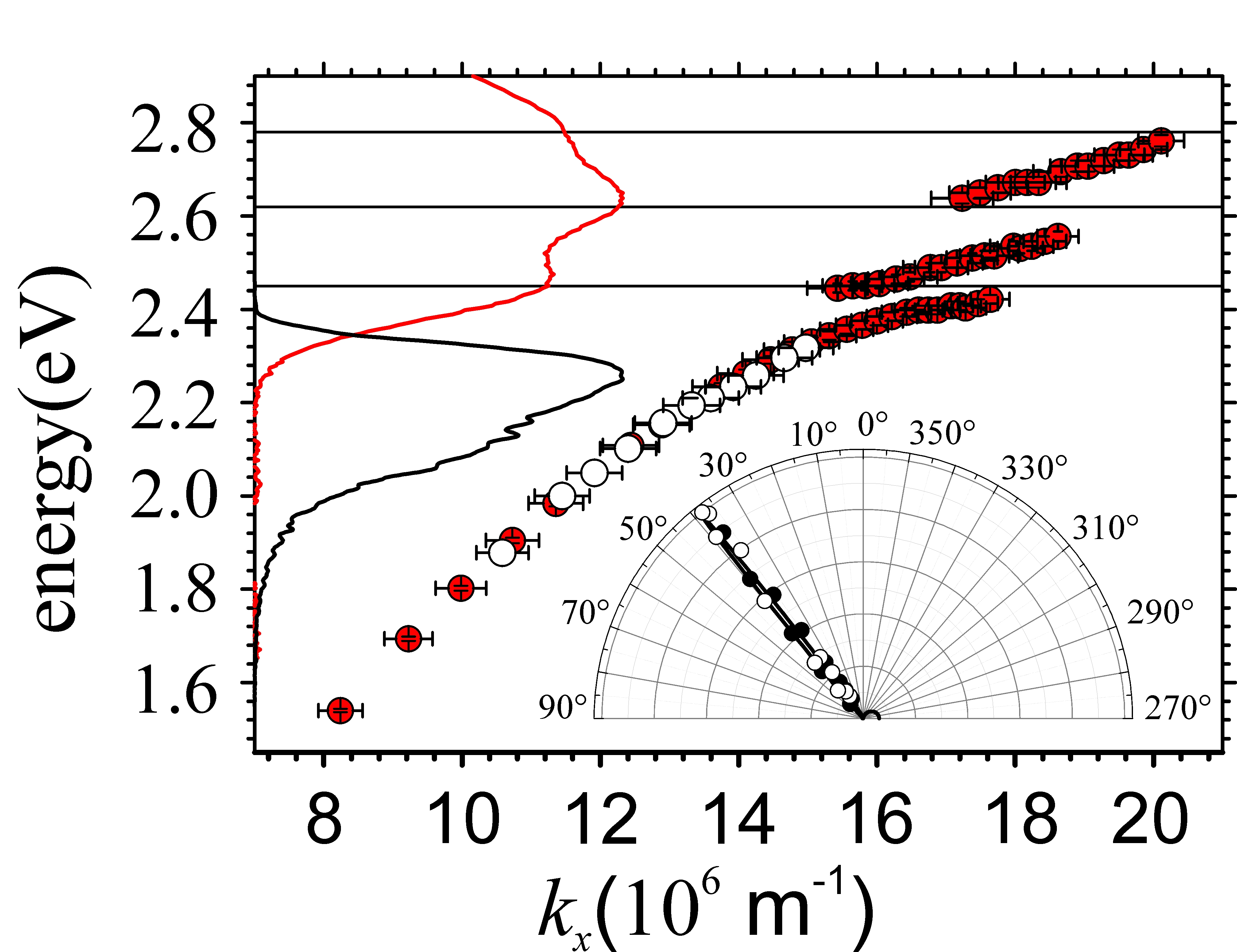
- Fig. 1: Dispersion relations of glass/ 50 nm silver/ 50 nm β-carotene structure obtained from the attenuated total reflectance (red circles) and surface plasmon coupled emission (empty circles) measurements are shown together with β-carotene absorption (red curve) and fluorescence (black curve) spectra. Inset shows measured radiative pattern of the same structure excited with two excitation wavelengths (solid and empty circles) and calculated one (black curve).
[1] S. Baieva, J.A. Ihalainen, J.J. Toppari, J. Chem. Phys. 138 (2013) 044707.
Nanotheranostics@CIGMH, Department of Life Sciences, Faculdade de Ciências e Tecnologia, Universidade Nova de Lisboa, Caparica, Portugal
Gold nanoparticles have attracted considerable attention in molecular recognition applications due to their simplicity and versatility, becoming a critical component in the development of nanotechnology-based approaches for in vitro diagnostics assays. Special attention has been paid to the development of assays and biosensing platforms capable of specific identification of nucleic acid sequences that can be integrated into genome screening strategies and identification of sequence polymorphisms associated to relevant phenotypes, or identification of pathogens1. AuNPs have been extensively used because of their ease of synthesis and unique optical properties with their typical bright red color in colloidal solutions associated with the localised surface plasmon resonance band (LSPR). LSPR is dependent on size, composition, shape and inter-particle distance. Another remarkable property is the easiness of chemical functionalization via the use of thiol-ligands that form quasi-covalent bonds between any given biomolecules (used as probe) and the gold surface, such as oligonucleotides2, rendering them suitable for application in bioassays.
We have developed a method based on the change of colour of a solution constituted by AuNPs functionalized with thiol-ssDNA mediated by a modification of the medium dieletric together by recognition of a target. The colour change occurs due to the decrease of the inter-particle distance that results in a red-shift of the typical LSPR and the concomitant change from red to blue. Here, I shall demonstrate the versatility of this system for the development of simple and robust nucleic acid detection assays: i) DNA – pathogen identification, SNP characterisation, mutation detection; ii) RNA without retrotranscription – quantification of gene expression and direct evaluation of fusion genes involved in cancer, miRNA detection; iii) integration in alloys for multi-colour detection.
Funding by FCT/MEC [CIGMH (PEst-OE/SAU/UI0009/2011); PTDC/QUI-QUI/112597/2009; PTDC/CTM-NAN/109877/2009; PTDC/BBB-NAN/1812/2012] is gratefully acknowledged.
[1] P. Baptista P, etal. Anal Bioanal Chem 2008; 391:943-950.
[2] C. Mirkin etal. Nature 1996; 382:607-609
School of Physics and Astronomy, University of Exeter, Exeter, EX4 4QL, UK
We combine chiral plasmonic nanostructures with achiral dye molecules and measure the circular polarization dependence of the enhanced photoluminescence to probe the electromagnetic chirality in the near-field associated with the plasmonic nanostructures. Depending on the chiral symmetry of the near-fields, we find a distinct dissymmetry in the enhanced emission: the enhancement is greater for the handedness corresponding to the chirality of the near fields of the underlying plasmonic structures. This effect is reversed for the near fields of the enantiomeric structures and vanishes for the achiral near fields around achiral nanoparticles.
1 Humboldt-Universität zu Berlin, Department of Chemistry, Brook-Taylor-Str. 2, 12489 Berlin, Germany
2 BAM Federal Institute for Materials Research and Testing, Richard-Willstätter-Str. 11, 12489 Berlin. Germany
3 Centre for Biospectroscopy, School of Chemistry, Monash University, 3800 Clayton, Victoria, Australia
In analytics, surface-enhanced Raman scattering (SERS) became a powerful tool to gain information from biomolecules. By the use of noble metal nanoparticles as SERS substrates, high enhancement factors can be achieved. On this poster, we present SERS spectra of red blood cells and their components: hemoglobin and cell membranes. We report SERS spectra of Hb using silver nanoparticles at very small nanoparticle : Hb molecule ratios, that is, under conditions relevant for SERS-based nanotoxicity experiments with red blood cells at high sensitivity. We show that the structural information obtained in the experiment is highly dependent on the conditions under which the interaction of nanoparticles with Hb molecules takes place. An excitation wavelength in the near-infrared region (785 nm) is used to prevent cell damage. The SERS spectra from isolated hemoglobin and cell membranes enabled us to assign the spectral information from whole red blood cells. In addition to NIR-excitation, Raman scattering of isolated oxygenated hemoglobin was also excited at shorter wavelengths, and yielded different enhanced signals of porphyrin and globin due to pre-/resonant enhancement. In contrast to previous work, where information about the hemoglobin protein structure is mainly obtained in resonant Raman experiments in the UV-range [1], in SERS we find globin bands as well as additional vibrations of the porphyrin, also at longer wavelengths. In addition to the information on hemoglobin structure, our results have important implications for our understanding of the interaction of red blood cells with nanoparticles [2].
We acknowledge funding by ERC starting grant No. 259432 (MULTIBIOPHOT) and the Australian Academy of science – scientific visits to Europe program.
[1] J. Kneipp, G. Balakrishnan, T. G. Spiro, The Journal of Physical Chemistry B 108 (2004) 15919-15927.
[2] D. Drescher, T. Büchner, D. McNaughton, Physical Chemistry Chemical Physics accepted (2013), DOI: 10.1039/C3CP43883J.
Department of Photonics, National Cheng Kung University, Tainan, Taiwan
Nanospherical-Lens Lithography (NLL) is an economic fabrication technique that is capable of fabricating nanodisk arrays that cover large area. It utilizes polystyrene nanospheres as focusing lenses to focus the incoming ultraviolet light and exposure the underlying photoresist layer. Photoresist hole arrays form after developing. Metal nanodisk arrays can be fabricated following metal evaporation and lifting-off process. Nanodisk arrays with diameter less than 100 nm and cover an area as large as 1 cm2 are being fabricated regularly using NLL in our research group.
In this study, NLL is used to fabricate nano-ellipse arrays by replacing the light source with a commercial ultraviolet lamp. The UV light from the lamp is propagating differently between the directions perpendicular and parallel to the lamp and therefore demonstrates different focusing behavior. We can control the orientation of the nano-ellipse since the long-axis is found to be parallel to the lamp direction. We also perform a throughout investigation on the effects of all the related parameters and nano-ellipse with aspect ratio of > 3 can be fabricated. The Localized Surface Plasmon Resonance (LSPR) of the nano-ellipse arrays is in infrared region when the long-axis length exceeds 1 m. Therefore, the fabricated nano-ellipse arrays are ideal platforms for surface-enhanced infrared absorption (SERIA). Nano-cross or nano-star arrays are also fabricated after double or triple exposure. We can also vary the exposure duration, the angle between exposures, and the sample location to fabricate more complicated metamaterials, such as T-, L-, U-, and H-shaped nanoparticle arrays, as shown in Fig. 1. More complicated patterns can be created by carefully designing the multiple exposure procedures. This proposed method is a powerful tool to fabricate multiple infrared metamaterials in large-scale, which will be very useful for various infrared metamaterial applications.
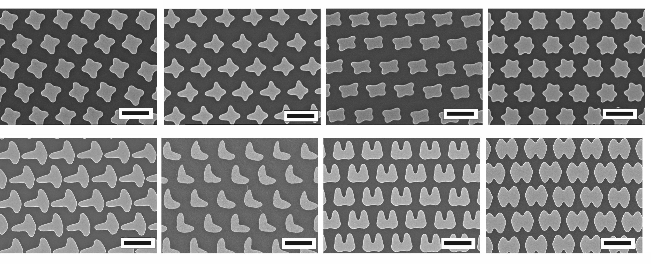
- Fig. 1: Various IR metamaterials fabricated with Nanospherical-Lens Lithography. Scale bar is 2 m.
The National Institute of Laser enhanced Science, Cairo University 12316 Giza, Egypt
Semiconductor quantum dots (QDs) are considered as a lately improved sector of nanomaterial with specific photophysical characteristics that increase the interest and potentiality of emerging what is known as fluorescent biosensors. For biosensing QD, they are advantageous over many of the conventional used protein-based fluorophores and organic dye that suffer from various chemical and photophysical liabilities. They include pH dependent, susceptibility to photo-bleaching, self-quenching at high concentrations, narrow absorption windows coupled to broad red-tailed emission spectra via small Stokes shifts, short-term aqueous stability and short excited state fluorescent lifetimes involve broad absorption spectra coupled to narrow size tunable photo fluorescent emissions. On the other hand QDs are characterized by having high quantum yields and unique resistance for both chemical degradation and photobleaching. The current research studies the synthesize of the CdSe core with a layer of wider band gap semiconductor such as CdTe QDs with different sizes for investigating their efficiency and sensitivity in adapting QDs for biosensing applications as generalized probes, in glycoproteins detection using laser induced florescence and fluorescence resonance energy transfer (FRET) - based sensing. The study as well focused on the selection of materials and the methodology for attaching biomolecules to the QDs that relies on direct interaction with the QD surface through dative thiol bonding of cysteine residues to the surface sulfurs.
[1] Kim, E.S.; Thomas, P.; Igor, L.M.; and Hedi, M. Sensors 6 (2006) 925-953.
[2] Giepmans, B.N.G.; Adams, S.R.; Ellisman, M.H.; Tsien, R.Y. Science 312 (2006) 217-224.
[3] Medintz, I.L.; Uyeda, H.T.; Goldman, E.R.; Mattoussi, H. Nature Materials 4 (2005) 435-446.
IPHT Jena, Albert Einstein Strasse 9, 07745 Jena, Germany
*Nanoscience Center, Department of Physics, P.O. Box 35, 40014 University of Jyväskylä, Finland
We demonstrate an excitation transfer along a fluorescently labeled dsDNA nanowire over a length of several micrometers. Launching of the excitation is done by exciting the localized surface plasmon mode of a 40 nm silver nanoparticle by 800 nm femtosecond laser pulses via two-photon absorption. The plasmonic energy is subsequently coupled to the nanowire and propagates along it inducing a bleaching of the dye. In situ as well as ex situ fluorescence microscopy was utilized to observe the phenomenon. In addition, transfer of the energy to another dye labeled nanoparticle over a separation of 5.7 µm was observed [1]. The observed phenomenon could be utilized in novel molecular systems providing communication method between the molecular devices.
Also, we show a new approach extending plasmonic lithography with the potential for a highly parallel nanofabrication with a higher level complexity based on nanoantenna effects combined with molecular nanowires. Thereby femtosecond laser pulse light is converted by silver nanoparticles into a high plasmonic excitation guided along attached DNA structures. An underlying poly (methyl methacrylate) (PMMA) layer acting as an electron sensitive resist is so structured along the former DNA position [2]. This apparently DNA guided effects leads to nanometer grooves reading even micrometer away from the excited nanoparticle, representing a novel effect of long range excitation transfer along DNA nanowires.
1 Nanobiophysics, MIRA Institute of Biomedical Technology and Technical Medicine,
2 Laboratory of Biophysics, Wageningen University, Wageningen, The Netherlands
3 The MacDiarmid Institute for Advanced Materials and Nanotechnology, School of Chemical and Physical Sciences, Victoria University of Wellington, Wellington, New Zealand
ield enhancement around noble metal nanoparticles can lead to enhancement of light-matter interactions with nearby molecules. While the enhanced fields can potentially enhance fluorescence from nearby molecules, in many cases quenching by the metal is dominant. While for single metal nanoparticles, fluorescence quenching is observed near the surface, and enhancement is observed only 5-10 nm farther aways, in gaps between particles, high fluorescence enhancement was theoretically predicted even at distances of 1nm and less.
We have recently shown1 that large fluorescence enhancement is possible from dyes co-aggregated with Ag nanoparticles (Ag-NPs). We seek to further our understanding of the interaction of dyes with the enhanced EM-field at hotspots between Ag-NPs. For a range of selected dyes, we have systematically measured the modification of the fluorophore excitation and emission spectra, the changes in fluorescent lifetime, and the effect of continuous aggregation on nanoparticle-enhanced fluorescence. The co-existence of high fluorescence enhancement (more than 100x) with a sharp decrease in the lifetime (from 3 ns to below 10ps) proves that the dyes experience giant field enhancement, which we believe is generated at hotspots between particles (see figure 1).2
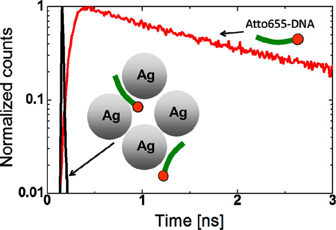
- Fig. 1: Observed fluorescence lifetime of Atto655 dye-labeled DNA in the absence and presence of silver nanoparticles aggregates
Additionally, we have looked at the enhancement of phosphorescence from a dye where fluorescence is spin forbidden. There we observe a very similar enhancement and lifetime modification behavior, however all the timescales are 3 orders of magnitude longer. We believe that combining phosphorescent dyes with plasmonic structures, even down to the single dye level, offers a convenient approach to better characterize plasmonic systems in detail.
Funding by the NWO (Veni grant No. 700.10.410) is gratefully acknowledged.
1. R. Gill and E. C. Le Ru, Phys. Chem. Chem. Phys., 2011, 13, 16366-16372.
2. R. Gill, L. Tian, W. R. C. Somerville, E. C. Le Ru, H. Van Amerongen and V. Subramaniam, J. Phys. Chem. C, 2012, 116, 16687-16693.
1 Humboldt-Universität zu Berlin, Department of Chemistry, Brook-Taylor-Str. 2, 12489 Berlin, Germany
2 BAM Federal Institute for Materials Research and Testing, Richard-Willstätter-Str. 11, 12489 Berlin. Germany
The Raman scattering of molecules which are adsorbed to plasmonic nanostructures is enhanced by several orders of magnitude because of the high local electromagnetic field in the proximity of the nanostructures. In surface-enhanced Raman-scattering (SERS), silver or gold nanoparticles are often used in suspensions. Diffusion and aggregation of these nanoparticles, however, lead to constraints in the reproducibility of the spectra, which is not beneficial in analytical applications.
Recently, we have reported that nanoparticles which are immobilized on glass surfaces by organosilanes show homogeneous SERS enhancement at the microscopic level which gives the possibility to use SERS as a tool for the quantification of analytes [1].
In order to investigate the potential of SERS microscopic imaging, we have created macroscopically defined regions, containing different analyte molecules, on two-dimensional arrays of immobilized silver nanoparticles and analyzed the microscopic distribution of different molecular species in mapping experiments. The obtained surfaces serve as a model system for the evaluation of SERS spectra from microstructured samples. Furthermore, the spatially resolved spectral information can give insights into the interaction of analyte mixtures with immobilized nanoparticles.
We acknowledge funding by ERC starting grant No. 259432 (MULTIBIOPHOT).
[1] V. Joseph, M. Gensler, S. Seifert, U. Gernert, J. P. Rabe, J. Kneipp, J. Phys. Chem. C 2012, 116, 6859-6865.
Department of Optical Science and Technology, The University of Tokushima, Tokushima 770-8506, Japan.
Interaction of lasers with gold nanoparticles (Au NPs) generates rich physics and chemistry represented by the particle's thermal expansion, shockwave generation, local heating and bubble formation, particle melting, particle evaporation, and plasma generation and particle fragmentation. [1] These processes are very complicated and their details and inter-relationship remains mostly concealed. Therefore, we attempted to understand both experimentally and computationally the course of events that accompany the excitation of relatively large, 60-100 nm in diameter, Au NPs, dispersed in aqueous solution or supported on glass surface.
Here we describe the fundamental aspects of Au NP interaction that leads to nanoscale energy deposition to the surroundings through light amplification and heat generation.[1] The laser-NP interaction also changes the particle's phase by electron and lattice heating. These results help us to understand the physical processes underlying photothermal therapy of malignant cells as well as nanofabrication involving noble metals.
Three examples are shown. (1)Laser-induced size reduction dependent on pulse duration.[2] We revealed that the mechanism of femtosecond laser-induced size reduction of Au NPs differs from that of nanosecond laser size reduction (Fig. 1). (2) Effect of high pressure on laser-induced size reduction.[3] By applying high pressures > 22.1 MPa, laser fluence-dependent size with a monodisperse particle distribution was obtained because the size reduction stopped at a given laser fluence regardless of pulse numbers (Fig. 2). (3) Application to nanofabrication. The laser irradiation of Au NPs fabricated nanoholes on a glass substrate (Fig. 3).
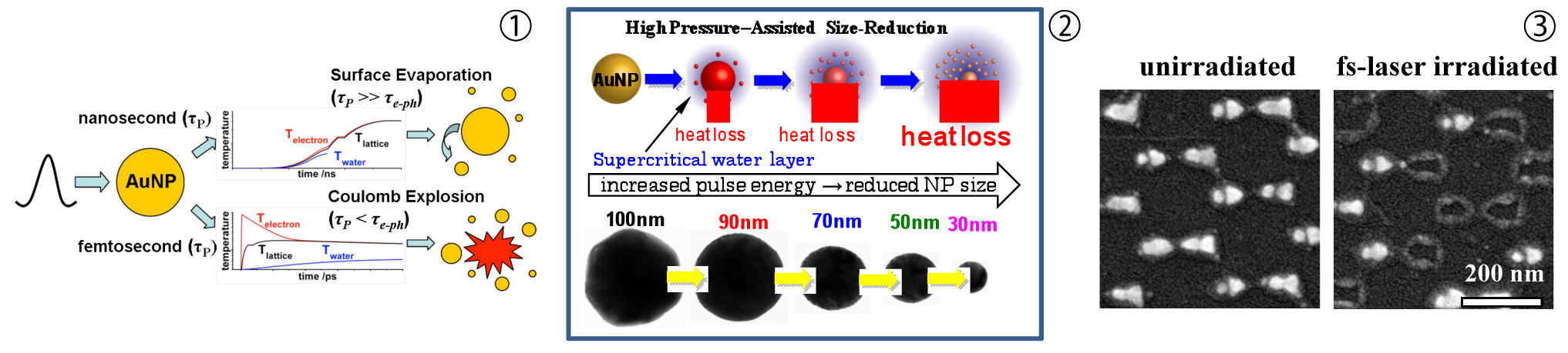
- Fig. 1: Temperature calculations explaining the difference of laser-induced size reduction of Au NPs between fs-laser and ns-laser excitations.
Fig. 2: Schematic illustration representing the nanosecond laser-induced size reduction of aqueous colloidal Au NPs at pressures > 22.1 MPa.
Fig. 3: Single-shot fs laser irradiation (: 800 nm) on patterned Au nanostructure (right) forms a replica on the surface (left).
[1] A. Plech et al., Laser Photon. Rev.,3 (2009) 435-451, S. Hashimoto et al., J. Photochem. Photobiol. C, 13 (2012) 28-41.
[2] D. Werner et al., J. Phys. Chem. C, 115 (2011) 5063-5072.
[3] D. Werner et al., Langmuir, 29 (2013) 1295-1302.
1 Institute of Physical Chemistry and Abbe-Center of Photonics, Friedrich-Schiller-University Jena, Helmholtzweg 4, 07743 Jena, Germany 2 IPHT - Institute of Photonic Technology, Albert-Einstein-Straße 9, 07745 Jena, Germany
The requirements for a detection method used in medical diagnostics and bioanalytics are high specificity, high sensitivity as well as the handling of small sample volumes. A suitable detection method, which provides high sensitivity and specificity, means surface enhanced Raman spectroscopy (SERS). An enhanced electromagnetic field is induced at the surface of metal nanostructures to amplify the Raman signal. The enhanced electromagnetic field affects molecules in close proximity of the surface resulting in an increase of the Raman signal intensity of these molecules up to 6 until 10 orders of magnitude [1]. Lately, the combination of SERS and microfluidic lab-on-a-chip (LOC) device becomes more and more of interest for the application in the field of bioanalytics [2]. LOC systems enable the handling of small sample volumes and the implementation of preparation procedure. Thus, LOC-SERS combines a high specific and sensitive detection method with a powerful tool for sample preparation procedures. Especially, for onsite and in-field application, the combination of SERS and LOC devices shows a high potential as analytical method [3-5]. The application of a liquid/liquid microsegmented flow avoids depositions at the channel walls. This ensures the measurement of reproducible SERS spectra, which is an important requirement for quantitative investigations [6]. Moreover, the Raman signal of the separation medium within the segmented flow LOC-SERS device is used for a wavenumber calibration [7].
In this contribution the great potential of SERS in a microfluidic device for quantitative analysis will be introduced by applying this technique as analytical tool for the characterization of the heteroaromate nicotine and its metabolite cotinine.
To analyze the SERS response, chemometric data evaluation is applied using the separation medium (n-tetradecane) for wavenumber calibration. This approach of employing the separation medium of a segmented flow as the wavenumber reference substance to calibrate the SERS spectra detected within the droplets allows the online-monitoring and the calibration of unwanted wavenumber fluctuations during the measurement due to ambient variations. Thus, a reliable and robust SERS-based measurement is achieved.
Acknowledgement: Funding the projects "QuantiSERS" and "Jenaer Biochip Initiative 2.0" within the framework "InnoProfile Transfer – Unternehmen Region" the Federal Ministry of Education and Research, Germany (BMBF) is gratefully acknowledged.
[1] Pettinger, B., Molecular Physics 2010, 108(16), 2039.
[2] Chen, J.X., Choo, J.B., Electrophoresis 2008, 29 (9), 1815.
[3] Ackermann, K. R., Henkel, T., Popp, J., Chemphyschem 2007, 8, 2665.
[4] März, A., Ackermann, K. R., Malsch, D., Bocklitz, T., et al., Journal Of Biophotonics 2009, 2, 232.
[5] Strehle, K. R., Cialla, D., Rosch, P., Henkel, T., et al., Analytical Chemistry 2007, 79, 154.
[6] März, A., Henkel, T., Cialla, D., Schmitt, M., Popp, J., Lab Chip 2011, 11, 3584.
1 Department of Physics , National Taiwan University, Taipei 10617 , Taiwan
2 Graduate Institute of Applied Physics , National Taiwan University, Taipei 10617 , Taiwan ,
3 Department of Physics , University of Massachusetts Boston, MA 02125 , USA
4 School of Electrical and Electronic Engineering , Nanyang Technological University , 639798, Singapore
5 Optoelectronics Research Centre and Centre for Photonic Metamaterials , University of Southampton, Southampton SO17 1BJ , UK
6 Research Center for Applied Sciences , Academia Sinica, Taipei 11529 , Taiwan
*Presenting author e-mail: ross6320@gmail.com
In the field of plasmonic metamaterials, the subwavelength metallic structures play a role similar to atoms in nature. The interference of their near-field coupling at plasmonic resonance leads to a plasmon induced transparency (PIT) that is analogous to the electromagnetically induced transparency (EIT) [1] of atomic systems. A sensitive control of the PIT is crucial to a range of potential applications such as slowing light and biosensor [2, 3]. So far, the PIT phenomena often arise from the electric resonance, such as an electric dipole state coupled to an electric quadrupole state. Here, we report the first three-dimensional photonic metamaterial consisting of an array of erected U-shape plasmonic gold nanostructures that exhibits PIT phenomenon with magnetic dipolar interaction between magnetic metamolecules rather than the electric interaction (see Fig. 1) [4]. We further demonstrate using a numerical simulation that the coupling between the different excited pathways at an intermediate resonant wavelength allows for a πphase shift resulting in a destructive interference. This work paves a promising approach to achieve magnetic plasmon devices.
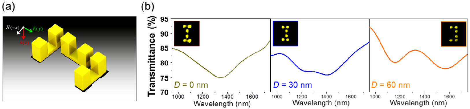
- Fig. 1: (a) Schematic diagram of a single SRR metamolecule unit cell. Each unit cell consisted of three erected gold SRRs with 600 nm period in both x and y directions. The optical properties of such metamolecule are studied in the case of y-polarized illumination. (b) The experimental transmittance spectra upon varying distance parameter D. All the insets show the top-view SEM images of the corresponding samples. excitations.
[1] K. J. Boller, A. Imamoglu, and S. E. Harris, Phys. Rev. Lett. 66, 2593-2596 (1991)
[2] Z. G. Dong, H. Liu , J. X. Cao, Li T, S. M. Wang, S. N. Zhu, X. Zhang . Appl. Phys. Lett. 97, 114101(2010)
[3] V . Yannopapas, E. Paspalakis , N. V. Vitanov. Phys. Rev. B 80, 035104 (2009)
[4] P.C. Wu, W. T. Chen, K. Y. Yang, C. T. Hsiao, G. Sun, A. Q. Liu, N. I. Zheludev and D. P. Tsai, Nanophotonics 1,131-138 (2012)
1 Graduate Institute of Applied Physics, National Taiwan University, Taipei 10617, Taiwan
2 Department of Physics, National Taiwan University, Taipei 10617, Taiwan
3 Research Center for Applied Sciences, Academia Sinica, Taipei 115, Taiwan
4 National Center for Theoretical Sciences at Taipei, National Taiwan University, Taipei 10617, Taiwan
5 Graduate Institute of Photonics and Optoelectronics, National Taiwan University, Taipei 10617, Taiwan
6 Department of Photonics, National Cheng Kung University, Tainan 701, Taiwan
7 School of Electrical and Electronic Engineering, Nanyang Technological University, Singapore
8 Institute of Opto-electronic Engineering, National Dong Hwa University, Hualien 97401, Taiwan
9 Physics Department, Fudan University, Shanghai 200433, China
*corresponding author: dptsai@phys.ntu.edu.tw
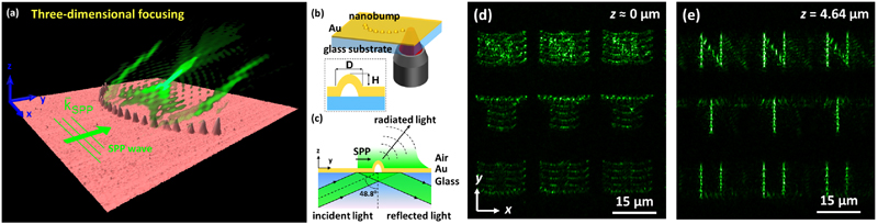
Using plasmonic nanostructures to manipulate the scattered light from the SPP and free-space impinging waves is demonstrated. The curved arrangement of Au nanobumps can scattered the surface plasmon waves and focused into spots in three-dimensional space. The light can be modulated into desired light patterns. For the free-space impinging waves, a gradient meta-surface supports broadband (750-900 nm) anomalous light reflections at ~850nm wavelength with high conversion efficiency (~80%) is realized.
Using nanostructures to manipulate surface plasmon polariton (SPP) plane waves is an important issue. Recently, three-dimensional focusing and diverging of SPP waves by a quarter circular structure composed of Au nanobumps were studied [1]. The Au nanobumps confer additional three-dimensional propagating wave vectors (kx, ky, kz) on SPP wave for departing from surface. In this work, we manipulate the scattering of SPP waves by various plasmonic structures composed of arranged nanobumps on a gold thin film [2]. Upon controlling the geometry of the plasmonic structures, the height, position, and pattern of scattered light can be modified as desired. It provides a simple and efficient way to project a specific light pattern into free space, and demonstrate the capability of three-dimensional light manipulation.
Gradient-index meta-surfaces were found to exhibit extraordinary light-manipulation abilities, governed by a generalized Snell's law with an additional parallel wave vector provided by the radiation phase gradient of the meta-surface. Recently, we showed that a new type of gradient meta-surface can convert a propagating wave to a surface wave with 100% efficiency provided that the phase gradient is large enough, and experimentally verified the idea in the microwave regime [3]. In this work, we designed and fabricated a gradient meta-surfaces working in visible region (~850nm) with broad-band functionality (750-900nm), and demonstrated by both experiments and numerical simulations that it can redirect an input light to a non-specular channel with high efficiency (~80%) [4].
In summary, we introduce two ways of light manipulation by the designed plasmonic nanostructures. Engineering the scattering properties of these nanostructures with the SPP or free-space impinging waves, the novel light-manipulation abilities can be achieved. Our results can lead to many practical applications, such as plasmonic beam splitters, three-dimensional plasmonic circuitry, holography, etc.
- Fig. 1: (a) Schematic diagram of a single SRR metamolecule unit cell. Each unit cell consisted of three erected gold SRRs with 600 nm period in both x and y directions. The optical properties of such metamolecule are studied in the case of y-polarized illumination. (b) The experimental transmittance spectra upon varying distance parameter D. All the insets show the top-view SEM images of the corresponding samples. excitations.

- Fig. 2: Schematic illustrations of (a) Geometry and working mechanism of our meta-surface. The designed sample consisting of Au nanorods (yellow) and a continuous Au film (yellow) separated by the MgF2 spacer (blue). (b) FDTD simulated scattered Ey field patterns of the gradient meta-surface under the normal incident of a y-polarized light with λ = 850 nm (c) Reflection phase of each structural unit within a super cell.
The authors acknowledge financial support from National Science Council, Taiwan under grant numbers 100-2923-M-002-007-MY3, 101-2112-M-002-023-, 101-2911-I-002-107 and 101-3113-P-002-021- respectively. They are also grateful to National Center for Theoretical Sciences, Taipei Office, Molecular Imaging Center of National Taiwan University, National Center for High-Performance Computing, Taiwan, and Research Center for Applied Sciences, Academia Sinica, Taiwan for their support.
[1] C. M. Chang, C. H. Chu, M. L. Tseng, Y.-W. Huang, H. W. Huang, B. H. Chen, D.-W. Huang, and D. P. Tsai, "Light manipulation by gold nanobumps," Plasmonics 7(3), 563–569 (2012)
[2] C. M. Chang, M. L. Tseng, B. H. Cheng, C. H. Chu, Y. Z. Ho, H. W. Huang, Y.-C. Lan, D.-W. Huang, A. Q. Liu, D. P. Tsai, "Three-Dimensional Plasmonic Micro Projector for Light Manipulation," Adv. Mater.25(8), 1118-1123 (2012)
[3] S. Sun, Q. He, S. Xiao, Q. Xu, X. Li, and L. Zhou, "Gradient-index meta-surface as a bridge linking propagating waves and surface waves," Nature Materials 11, 426 ( 2012)
[4] S. Sun, K.-Y. Yang, C.-M. Wang, T.-K. Juan, W. T. Chen, C. Y. Liao, Q. He, S. Xiao, W.-T. Kung, G.-Y. Guo, L. Zhou, and D. P. Tsai, "High-efficiency Broadband Anomalous Reflection of Light by Gradient Meta-surfaces," Nano Lett. 12, 6223-6229 (2012)
1 Graduate Institute of Applied Physics, National Taiwan University, Taipei 106, Taiwan
2 Optoelectronics Research Centre and Centre for Photonic Metamaterials, University of Southampton, Southampton SO17 1BJ, UK
3 Centre for Disruptive Photonic Technologies, Nanyang Technological University, Singapore 637371, Singapore5Instrument Technology
4 Department of Physics, National Taiwan University, Taipei 106, Taiwan
5 Research Center for Applied Sciences, Academia Sinica, Taipei 115, Taiwan
Toroidal shapes are often found in bio-molecules, viruses, proteins and fats, but only recently it was proved experimentally that toroidal structures can support exotic high-frequency electromagnetic excitations that are neither electric or magnetic multipoles. Such excitations, known as toroidal moments, could be playing an important role in enhancing inter-molecular interaction and energy transfer due to its higher electromagnetic energy confinement and weaker coupling to free space. The toroidal spectral responses of solenoid-like toroidal metamolecule integrated by four gold U-shaped split-ring resonators (SRRs) at optical frequencies are experimentally and numerically studied. Using a model toroidal metamaterial system, we offer an opportunity of creating the "toroidal" lasing spaser, a source of coherent optical radiation that is fueled by toroidal plasmonic oscillations in the nanostructure.
Different from the electric dipole and magnetic dipole, the toroidal multipoles are not included in the traditional multipole expansion [1]. Toroidal metamaterials were first theoretically proposed in 2007 [2]. The toroidal response results in strong dependence on the dielectric permittivity, which can make negative index of refraction [2] and rotate polarization state of light [3]. In 2010, the toroidal metamaterials consisted by four 3D resonant split metallic wire loops show toroidal response in microwave [4].
In this paper, we experimentally and numerically study the optical response of toroidal metamolecule. Figure 1(a) shows the SEM image of three-dimensional resonant rings fabricated by double exposure e-beam lithographic process with 150 nm length, 45 nm width and 60 nm height. The plasmonic responses of solenoid-like toroidal metamolecule integrated by four gold U-shaped SRRs is studied by solving 3D Maxwell equation with the finite-element method. The toroidal metamolecule is illuminated by linearly polarized light with magnetic field Hx passing through the SRRs, which can support a strong magnetic dipole moments on each single SRR. Figures 1(b) and 1(c) show the magnetic energy density and field lines at magnetic and toroidal resonances, respectively. Incident light induced magnetic dipoles point in the same direction produced magnetic resonance. In contrast, four magnetic dipoles form a head-to-tail configuration which concentrates toroidal resonance [5].We also propose a lasing spaser that is fueled by previously overlooked toroidal dipolar mode of plasmonic coherent oscillations. Figure 1 (d) shows the schematic diagram of the plasmonic toroidal lasing spaser. Such a high Q-factor of toroidal metamolecule with a narrow bandwidth of gain has applications, such as single pass amplifier which the certain frequency of incident wave can be amplified up to 65 dB [6].
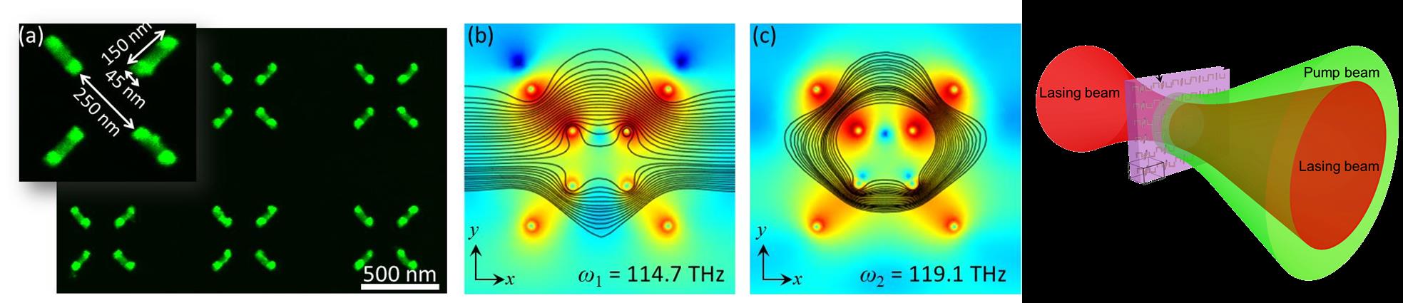
- Fig. 1: (a) SEM image of three-dimensional gold resonant split rings. Distribution of magnetic field lines (black streamline) and magnetic energy (color map, in logarithm scale) of magnetic (b) and toroidal (c) resonances. (d) Schematic diagram of the plasmonic toroidal lasing spaser. The structure comprises an amplifying medium slab (purple) supporting an array of plasmonic toroidal metamolecules.
[1] V. M. Dubovik, and V. V. Tugushev, Phys. Rep. 187, 145 (1990).
[2] K. Marinov, A. D. Boardman, V. A. Fedotov, and N. Zheludev, N. J. Phys. 9, 324 (2007).
[3] N. Papasimakis, V. A. Fedotov, K. Marinov, and N. I. Zheludev, Phys. Rev. Lett. 103, 093901 (2009).
[4] T. Kaelberer, V. A. Fedotov, N. Papasimakis, D. P. Tsai, and N. I. Zheludev, Science 330, 1510-1512 (2010).
[5] Y.-W. Huang, W. T. Chen, P. C. Wu, V. Fedotov, V. Savinov, Y. Z. Ho, Y. F. Chau, N. I. Zheludev, and D. P. Tsai, Opt. Express 20, 1760-1768 (2012).
[6] Y.-W. Huang, W. T. Chen, P. C. Wu, V. A. Fedotov, N. I. Zheludev, D. P. Tsai, Scientific Reports 3, 1237 (2013).
Institute of Physics and Center for Interdisciplinary Nanostructure Science and Technology, University of Kassel, Germany
We start this presentation with a contribution to the long standing discussion, which addresses the origins of SERS enhancement, i.e. chemical enhancement or electromagnetic enhancement. Usually high enhancement factors are attributed to high electromagnetic fields. However, very often the corresponding SERS spectra show clear features of chemical enhancement. This contradiction is mostly ignored when explaining SERS enhancements in the order of 1010 or higher. The reason is that present theories predict only for electromagnetic enhancements sufficiently high enhancement factors to explain the strong SERS signals. To solve the contradiction, we propose a new scheme in which the excitation of plasmon resonances supports the chemical enhancement via chemical interface damping [1]. We demonstrate that high SERS signals can also be explained by chemical enhancement.
Regardless of the exact mechanism of SERS enhancement, trace detection of pollutant molecules in water is an important task in environmental science. In particular, polycyclic aromatic hydrocarbons (PAHs) are toxic to biota even in very low concentrations and bioaccumulate in aquatic organisms. Thus, for routine detection of PAHs in water a reliable, molecule specific, and simple technology is desired, which is capable to measure small molecular concentrations. Along these lines, we demonstrate in the second part of this presentation that trace detection becomes feasible by combining surfaced enhance Raman spectroscopy (SERS) with shifted excitation difference Raman spectroscopy (SERDS) and using tailored noble metal nanoparticle ensembles as SERS substrates. While SERDS improves the signal to noise ratio significantly, the tailored nanoparticle ensembles yield sufficient high SERS signals. With this set up we have measured in aqueous solutions PAH concentrations as low as 4 nmol/l for flouranthene and 6 nmol/l for pyrene with a response time below 10 minutes. However, the calculated limit of detection for both molecules is in the range of only 2 nmol/l, i.e. sufficient to detect the maximum allowable concentration of PAHs in water, as defined by the European Quality Standard. Hence, we have developed an ideal system that can be used as an alarm sensor for PAHs in water.
[1] F.H., J. Raman Spectr., submitted
[2] Y. H. Kwon, R. Ossig, F.H., H.-D. Kronfeldt, J. Raman Spec., DOI 10.1002/jrs.4093 (2012)
[3] R. Ossig, A. Kolomijeca, Y.-H. Kwon, F.H., H.-D. Kronfeldt, J. Raman Spec., accepted.
1 University of Cambridge, Sector for Biological and Soft Systems, Cavendish Laboratory, 19 JJ Thompson Avenue, CB3 0HE, Cambridge, UK
2 University of Cambridge, Centre for Brain Repair, Forvie Site, Robinson Way, CB2 0PY, Cambridge, UK
3 University of Southampton, Department of Chemistry, Faculty of Natural & Environmental Sciences, Highfield, SO17 1BJ, Southampton, UK
The segregation of closely related cell phenotypes by non-invasive imaging techniques like optical microscopy is currently impossible both in vitro and in vivo. Our approach employs surface-enhanced Raman spectroscopy (SERS) for cellular imaging to address this problem. Raman scattering is a vibrational spectroscopic technique which gives a molecular fingerprint of detected molecules. Though, Raman scattering is a very weak process, significant enhancement can be achieved by bringing metal nanoparticles into close vicinity of the molecule of interest which is known as SERS. In contrast to conventional spectroscopic methods, SERS is label-free, non-invasive and does not need fluorescent markers for imaging molecules inside cells.
We have used spherical gold nanoparticles as intracellular SERS transducers in neuronal cells (SH-SY5Y). Aggregated nanoparticles inside cells allow for SERS imaging revealing a chemical map of the cell. Data achieved in only a single scan contains information about the intracellular distribution of various biochemical compounds such as lipids and proteins.
Furthermore, we are able to distinguish between progenitor and differentiated neuronal cells using cytoplasmic and nuclear SERS spectra. Functionalisation of gold nanoparticles with the nuclear localisation signal peptide sequence [1] was used to target the cell nucleus. As the relative amount of nuclear DNA/ RNA varies with the state of cellular differentiation, SERS spectra from the nucleus allow for distinction between progenitor and differentiated cells. Additionally, we found that the level of protein increases during differentiation. Our approach allows for segregation of cell types as well as characterisation of molecular changes during cellular differentiation.
Thus, targeted SERS nanoparticles provide an appropriate probe for intracellular imaging by preserving the integrity of the biological system and showing many advantageous over conventional spectroscopic techniques.
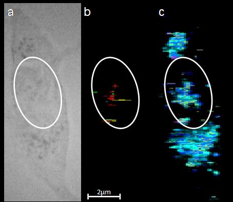
- Fig. 1: Bright field image (A) and SERS map of a differentiated SH-SY5Y cell highlighting the intracellular distribution of DNA/RNA (B) and proteins (C)
We gratefully acknowledge EPSRC funding support (EP/H028757/1; EP/G060649/1) for this work.
[1] A. G. Tkachenko, H. Xie, D. Coleman, W. Glomm, J. Ryan, M. F. Anderson, S. Franzen, D. L. Feldheim, Journal of the American Chemical Society 125, 4700-4701, 2003.
Nanoscience Center, 1 Department of Physics and 2 Department of Chemistry, P.O.Box 35 (YN)
FI-40014 University of Jyväskylä, Finland
tommi.isoniemi@jyu.fi
In carbon materials, actual optical plasmonics have this far been restricted to graphene, but the experiments we have performed [1] provide novel evidence of plasmonic functionality in a different form of pure carbon. We demonstrate optical resonances in thin films of single walled carbon nanotubes (SWCNTs) with a highly enriched (98 %) proportion of metallic chiralities. These resonances are measured in the Kretschmann configuration, and can be seen as intensity dips of up to 90 % in reflection spectra at 400 and 700 nm (Fig. 1). Unexpectedly, they are only visible when the sample is excited with s-polarized light, the opposite of surface plasmon polaritons on thin metal films. The resonances are dispersive and intense only when the layer thickness is close to 100 nm, implying that a collective excitation might be responsible for the resonance. They are also sensitive to the dielectric environment, clearly distinguishing the data from normal total internal reflection absorption spectra. The length of the CNTs seems to be irrelevant, ruling out localized surface plasmon resonance, and increasing the amount of amorphous carbon only decreases the intensity of the resonance. Although additional experimental and theoretical studies are needed to confirm the underlying mechanism, a magnetic plasmon resonance due to intertube effects, possibly within bundles, is a possible explanation. A probable coupling to excitons can also be pointed out, as the resonances are found close to M11 and M22 transition energies of the SWCNTs. If the fundamental reason for the observed phenomenon is connected to a magnetic resonance, metallic SWCNTs might find applications in plasmonic metamaterials.
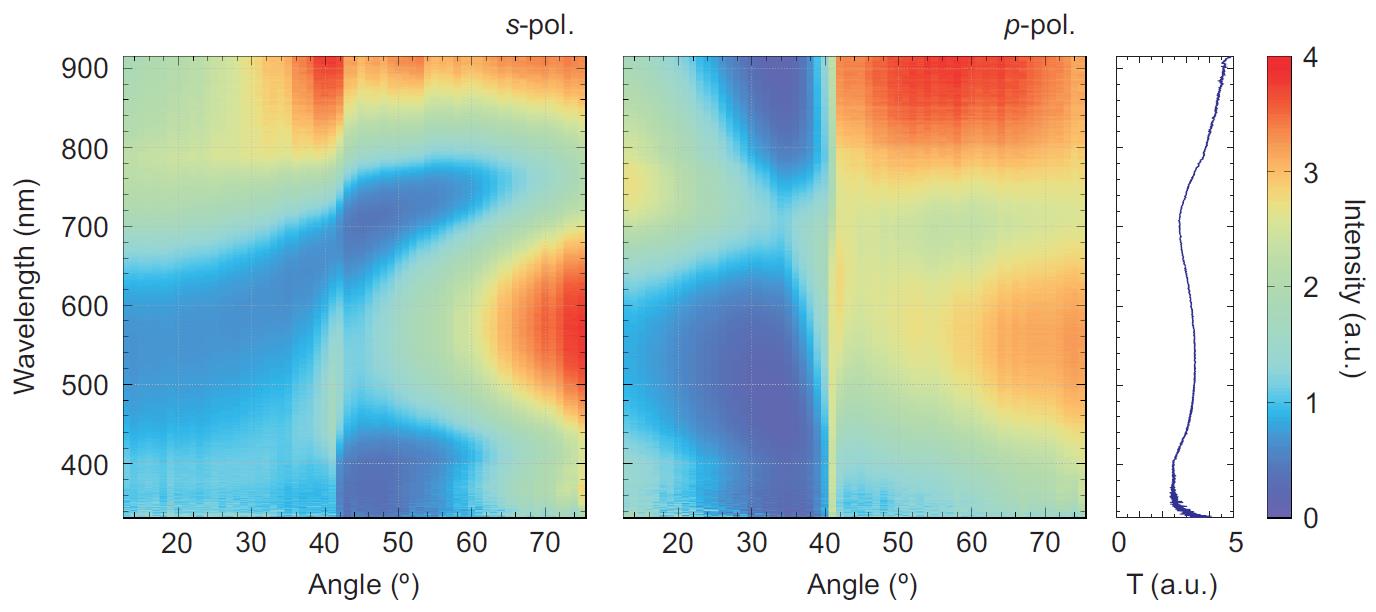
- Fig. 1: Kretschmann reflection spectra at s- and p-polarizations of an approximately 100 nm thick metallic SWCNT film on a glass substrate. Total internal reflection at angles greater than 41º. Transmission spectrum of the sample at right.
[1] Isoniemi, T., Johansson, A., Toppari, J.J. & Kunttu, H. 2013 Collective optical resonances in networks of metallic carbon nanotubes. Submitted.
Institute of Photonic Technology, Albert-Einstein-Str. 9, 07745 Jena, Germany
Nanoholes and nanoparticles have both found their way in todays bio-analytic. Here we present a hybrid nanostructure consisting of non-plasmonic nanoholes and plasmonic nanoholes. The study of this structure reveals a great potential for bio-analytical applications. The investigation was performed with ultramicroscopic methods like AFM and SEM for the characterization of the geometric properties of the hybrid nanostructure. The more important study was done by the micro-spectroscopic measurement of the optical behaviour of the nanostructure in the transmitted light mode with a optical microscope. These measurements show on the one hand the change of the optical properties of the nanoholes in the case a nanoparticle is present and one the other hand the optical change after the immobilization of bio-molecules is also visible. With the knowledge of these properties the extraction of spectral features from microscope images is possible. With an automated image processing for the hybrid nanostructure enables the high throughput of the hybrid nanostructure.[1] The image processing and the micro-spectral measurements show in combination with biological test systems a great potential high sensitive bio sensoric measurments.[2]
[1] Jahr, N. et. al., International Journal of Environmental Analytical Chemistry 93(2) (2013) 140-151
[2] Jahr, N. et. al., The Journal of Physical Chemistry C 117(15) (2013) 7751-7756
Institute of Photonic Technology, Albert-Einstein-Str. 9, 07745 Jena, Germany
Noble metal nanoparticles are dealt as promising transducers for the detection of biomolecules. Their application as biosensors are founded on their special optical properties called localized surface plasmon resonance (LSPR). The sensing principle is bases upon the detection of the refractive index change surrounding the nanoparticles, which is measured by the shift of the plasmonic peak.
The spectral shift can be determined by nanoparticles densely adsorbed as layer on glass measured as ensemble (eLSPR) or at the single nanoparticle level (sLSPR). We report about detection systems for both cases in-situ and compare their sensitivity for future bioanalytical applications. As biological model, ultra-thin (few nm thick) polymer layers were sequentially adsorbed on the nanoparticles by layer-by-layer deposition technique. The surface sensitivity and the volume sensitivity of different gold and silver nanoparticles e.g. spheres, peanuts, or prisms with eLSPR and sLSPR will be presented. Finally the results are compared to simulations and the established SPR technique (Figure 1).
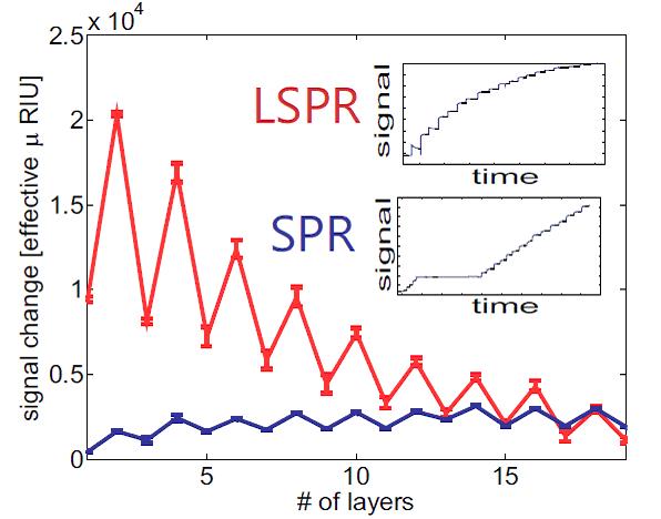
- Fig. 1: SPR and eLSPR sensitivity for each adsorbing polymer-layer on 80nm gold nanoparticles (red) and on gold surface (blue)
[1] Szunerits S.and Boukherroub R. (2012) Sensing using localised surface plasmon resonance sensors. Chem.Commun.,48 , 8999-9010.
[2] Sepúlveda B., et al. (2009). LSPR-based nanobiosensors. Nano Today Vol. 4, 244-251.
[3] Csáki A., et al. (2009). Plasmonic Nanoparticles – Noble Material for Sensoric Applications. Nova Science Publishers, Inc., ISBN 978-1-61668-009-1.
1 Institute of Solid State Physics, Friedrich-Schiller-University Jena, Helmholtzweg 5, D-07743 Jena, Germany
2 Institute of Photonic Technology (IPHT), Albert-Einstein-Str. 9, D-07745 Jena, Germany
The authors present a novel in-situ method of fabricating single-crystalline gold nanoparticles by self-organization using two different matrices (YBa2Cu3O7-δ and SrTiO3) grown by a pulsed laser deposition technique. Thereby a thin gold layer self assembles into crystalline nanoparticles due to the elevated temperatures during the matrix deposition. Through a variation of the initial seed layer thickness we are able to influence the size and the distribution of the particles [1]. Furthermore, by using a thin film matrix induced self-assembly of nanoparticles one gets the opportunity of controlling the nanoparticle shape. The interaction between growing thin film matrix and Au nanoparticles allows fabricating anisotropic particles which are highly attractive for the realization of photonic applications [2, 3].
As one might have to extract the nanoparticles or at least theirs tips from the surrounding matrix material to realize photonic applications we will also present our findings concerning the matrix dissolution. Furthermore, several possibilities will be given to grow nanoparticles only at well-defined positions on the substrate. A spectral characterization of particles will be presented based on microspectroscopy.
C. Katzer gratefully acknowledges financial support by the Landesgraduiertenförderung Thüringen.
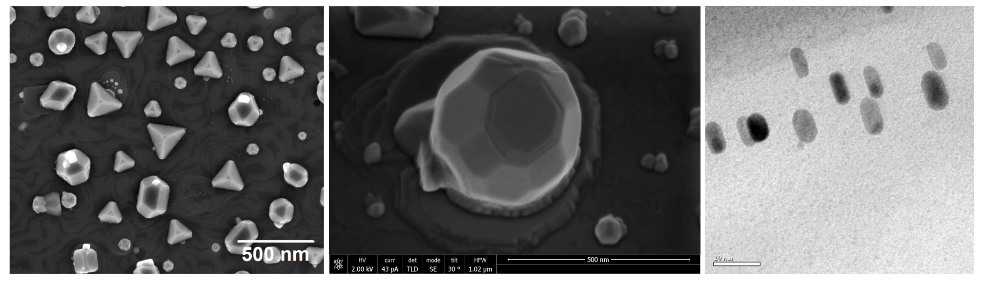
- Fig. 1: Au nanoparticles of different shapes and sizes, grown by a matrix induced self-assembly.
[1] V. Grosse, S. Engmann, F. Schmidl, A. Undisz, M. Rettenmayr and P. Seidel, phys. stat. solidi RRL 4 (2010), 97
[2] C. Katzer, V. Grosse, F. Schmidl, P. Michalowski, G. Schmidl, R. Mueller, J. Dellith, C. Schmidt, J. Jatschka and W. Fritzsche, J. Nanopart. Res.14 (2012), 1285
[3] S. Christke,C. Katzer, V. Grosse, F. Schmidl, G. Schmidl, W. Fritzsche, J. Petschulat, T. Pertsch and M. Rettenmayr, Opt. Mater.
Express 1 (2011), 890–897
Biomedical Diagnostics Institute, School of Physical Sciences, Dublin City University, Glasnevin, Dublin 9, Ireland1
Microbiology, School of Natural Sciences, National University of Ireland, Galway, Ireland2
Molecular Diagnostics Research Group, National Centre for Biomedical Engineering Science, National University of Ireland, Galway, Ireland3
Tyndall National Institute, Lee Maltings, University College, Cork, Ireland4
The field of plasmonics is a rapidly growing area of research and holds much promise for the improvement of biomedical diagnostics. Fluorescence technology poses a viable approach to the development of biosensors capable of the early detection of disease, but improvements in sensitivity are needed. Of particular interest is metal enhanced fluorescence (MEF),1 which could offer a route to more sensitive biosensor systems. In fact, our group have previously shown a 66 fold improvement in the limit of detection for a human IgG assay on a silver nanoparticle functionalised polystyrene substrate when compared with the assay in the absence of the nanoparticles.2 Here we extend this approach for sensitive DNA detection.
Glass substrates were functionalised with polyelectrolyte layers (PELs) via layer-by-layer self assembly. Silver nanoparticles (~150 nm) were uniformly and reproducibly deposited on the polyelectrolyte functionalised glass to form our detection platform. The substrates were characterised by UV-Vis spectroscopy, SEM and contact angle measurements. Thiol-terminated oligonucleotides (20 mer) were immobilised on the silver surface as capture probes. Hybridisation of Cy5-labelled target strands (80 mer), with both complementary and non-complementary probes, were performed to assess the performance of our platform. Successful and selective hybridisation was observed within 15 minutes. Cy5 fluorescence of hybridised target was detected down to 200 pM. A comparison of our platform with a commercial glass platform shows an increased fluorescence signal and enhanced limit of detection capabilities for the silver platform.
[1] C.D. Geddes (Editor), Metal-Enhanced Fluorescence; Wiley, (2010)
[2] R. Nooney, A. Clifford, X. LeGuevel, O. Stranik, C. McDonagh, B.D. MacCraith, Anal. Bioanal. Chem. 396 (2010), 1127-1134
1 Humboldt-Universität zu Berlin, Department of Chemistry, Brook-Taylor-Str. 2, 12489 Berlin, Germany and BAM Federal Institute for Materials Research and Testing, Richard-Willstätter-Str. 11, 12489 Berlin. Germany
2 Technical University of Denmark, Department of Chemistry, Kemitorvet 207, 2800 Kgs.Lyngby, Denmark 3 Technical University Berlin, ZELMI, Straße des 17. Juni 135, 10623 Berlin
Immobilization using organosilanes allows the generation of surfaces containing different types of nanoparticles at the same time. We have recently investigated such surfaces and determined SERS enhancement factors and their distribution at the microscopic level as a function of nanoscopic and plasmonic properties [1]. The concept of silane-immobilization of gold and silver nanostructures as SERS active substrates can be extended to generate mixed-nanoparticle surfaces with new surface functionalities for application in analytical sciences and in catalysis research. We have simultaneously immobilized separate gold and platinum nanoparticles on a glass surface and used them to map the kinetics of a catalytic reaction [2]. This is different from previously reported composite nanostructures of materials with plasmonic and catalytic properties. The proximity of both types of nanoparticles enables interaction of the molecules with the platinum nanoparticles whilst they reside in the local optical fields provided by the localized surface plasmons of the gold nanoparticles. The approach enables us to compare reaction rate constants of different catalytic systems and the underlying kinetics such as the formation of the active species independent of the optical absorption properties of the reaction products and / or the catalysts.
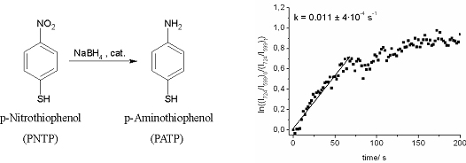
- Fig. 1: Determination of the rate constants for the reduction of nitrothiophenol with sodium borohydride (left) in the presence of Pt nanoparticles in SERS spectra obtained with APTES-immobilized gold nanoparticles.
[1] V. Joseph, M. Gensler, S. Seifert, U. Gernert, J. P. Rabe, J. Kneipp, J. Phys. Chem. C 116 (2012), 6859-6865.
[2] V. Joseph, Ch. Engelbrekt, J.Zhang, U. Gernert, J. Ulstrup, and J. Kneipp, Angewandte Chemie- International Edition 51 (2012) 7592–7596
Institute of Photonic Technology, Albert-Einstein-Straße 9, 07745 Jena, Germany
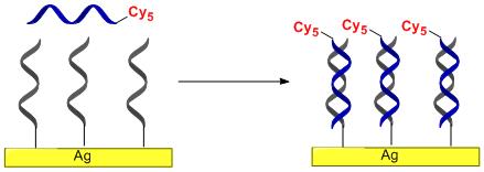
DNA-Origami has become to one of the most noted methods in DNA-Nanotechnology. With this bottom up technic almost any nanoscale DNA shape can easily formed by molecular self-assembly [1][3] and can be arranged to bigger 2D structures [2][4].
Metal nanoparticle structures are interesting for their optical properties caused by plasmonic effects. So in theory an accurately defined nanoparticle pattern could function as a "plasmonic fiber" or as an optical amplifier [5]. DNA-Origami structures can serve as nano-breadboards for the defined positioning of Biomolecules, single fluorophore dyes and metal nanoparticles with a spatial resolution of roughly 6 nm.
Here the gold nanoparticles were covered with thiol-modified staple strands and incubated with DNA-Origami rectangles [1] without the corresponding staple strands. This method seems promising to achieve not only an one-pot annealing for DNA structures but also an one-pot solution for DNA-nanoparticle shapes and pattern by skipping the hybridisation step on sticky ends.
We also present experiments for the defined metallization of 2D-DNA-Origami structures using gold-ions in solution. An easy and reliable method for the metallization of DNA-superstructures would be a step toward novel applications for these superstructures for nanophotonics or nano-circuitry.
- Fig. 1: A: spezific combination of four DNA-Origami labelled with DNA-hairpins ([2] modified) B: precise positioning of gold nanoparticles (15nm) on DNA-Origami
[1] P. Rothemund (2006). "Folding DNA to create nanoscale shapes and patterns". Nature 440 (7082): 297–302.
[2] S.Woo et al. (2011)."Programmable molecular recognition based on the geometry of DNA nanostructures" Nature Chemistry (VOL 3) :620-627
[3] Bryan Wei et al. (2012)."Complex shapes self-assembled from single-stranded DNA tiles", Nature Volume: 485, Pages: 623–626
[4] Arivazhagan Rajendran et al. (2011). "Programmed Two-Dimensional Self-Assembly of Multiple DNA Origami Jigsaw Pieces", ACSNANO VOL. 5 : 665–671
[5] S.Bidault et al. (2008)."Plasmon-Based Nanolenses Assembled on a Well-Defined DNA Template", J. Am. Chem. Soc.
1 Laboratoire CSPBAT-UMR7244, UFR Santé Médecine Biologie Humaine-Université Paris13, 74 rue Marcel Cachin, 93017 Bobigny, France, marc.lamydelachapelle@univ-paris13.fr
2 Biofunctional Nanomaterials Department, CIC biomaGUNE, Parque Tecnológico de San Sebastian, Donostia, San Sebastian, Spain
3 INSERM U698, Bioingénierie cardiovasculaire, Université Paris 13, 74 rue Marcel Cachin, 93017 Bobigny, France
4 Université de technologie de Troyes, Laboratoire de Nanotechnologie et d'instrumentation Optique, Institut Charles Delaunay, FRE 2848, 12 rue Marie Curie, 10010 Troyes, France
5 Istituto Italiano di Tecnologia (IIT). Via Morego, 30 16163 Genova, Italy.
The development of reliable, sensitive and specific biosensors is a very active research field. Among all the technique, the Surface Enhance Raman Scaterring (SERS) is one of most sensitive way to detect protein [1]. It has been widely used for ultrasensitive chemical analysis down to single molecule detection. Its field of applications now includes chemical-biochemical analysis, nanostructure characterization and biomedical applications. In this work, we present the SERS detection of specific proteins and more especially specific disease biomarkers in serum, using a functionalizion layer.
To investigate the protein detection, the arrays of metallic nanoparticles were made by electron-beam lithography (EBL) to control the Localized Surface Plasmon Resonance (LSPR) position in order to obtain the best enhancement and optimise the SERS signal [2-3]. The nanoparticles were in gold with different shapes: cylinders and rods. The optimization of plasmonic nanostructures to improve their sensing properties such as their sensitivity and their easy manipulation is of first importance in order to develop highly sensitive sensor. The key point is then the optimization of the localized surface plasmon resonance (LSPR) properties especially for surface-enhanced Raman scattering (SERS). Several aspects can be considered in order to optimize the sensing performance: size and shape of the nanoparticles, nanoparticle coupling, molecular adhesion layer between gold nanostructures and glass…[4] By controlling all these aspects, we are able to produce a highly sensitive sensor. To be specific, the surface was functionalized with aptamer or anti-bodies to catch selectively the targeted protein. We have determined the sensor characterisrics such as its detection limits and its selectivity. We have determined that such sensor could be highly sensitive by reaching some detection limits lowest than the pico-molar.
In conclusion, we report that we can detect the disease biomarkers using the LSPR and the SERS. Moreover, we demonstrate that the functionalization surface assure the specificity of the biosensor and allow to detect the target protein in the serum.
The authors want to acknowledge the Nanoantenna collaborative European project (HEALTH-F5-2009-241818) for financial support.
[1] K. Kneipp, Y. Wang, H. Kneipp, L. Perelman, I. Itzkan, R. Dasari, and M. Feld, Phys. Rev. Lett. 78/9 (1997) 1667-1670
[2] J. Grand, M. Lamy de la Chapelle, J.-L. Bijeon, P.-M. Adam, A. Vial, and P. Royer, Phys. Rev. B 7, (2005) 033407, 2005.
[3] N. Guillot , H. Shen , B. Fremaux , O. Péron , E. Rinnert , T. Toury, M. Lamy de la Chapelle, Applied. Phys. Lett. 97 (2010) 023113
[4] H. Shen , N. Guillot, J. Rouxel, M. Lamy de la Chapelle, T. Toury, Optics express 20(19) (2012) 21278
a Bionand, Nanomedicine centre, Technological Park of Andalucia, Malaga-Spain
b Institute for Applied Materials, KIT-Campus North, Karlsruhe, Germany
c Division of Pharmaceutical Technology, Faculty of Pharmacy, Helsinki, Finland
d Department of Biophysical Chemistry, Saarland University, Saarbrucken, Germany
e Pharmaceutical Nanotechnology, Saarland University, Saarbrucken, Germany.
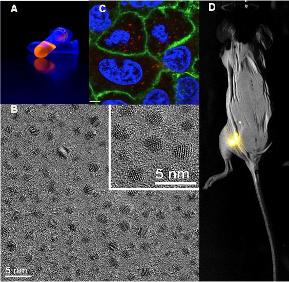
Noble metal nanoclusters (NCs) have stimulated extensive interest in the last five years due to their photophysical properties and their applications in nanomedicine. Several groups have studied the origin of the fluorescence of NCs and their structure–property inside different cavities such as polymers [2], dendrimer [3] and proteins [4]. Few-atom nanoclusters differ from gold or silver nanoparticles in that they can be highly fluorescent, do not support a surface plasmon, and do not have the metallic and bulk-like properties of nanoparticles/nanocomposites [5]. This fluorescence is likely due to the transition of molecule-like electronic levels when subnanometer sizes are smaller than the Fermi wavelength (i.e. < 1 nm). Noble metal (Ag, Au, Pt) NCs were prepared in wet chemistry in biological templates: proteins (Bovine Serum Albumin, human Transferrin) and peptide (Glutathione), which act as reducing agent and stabilizer. Spectroscopic investigations indicate a strong relation between the oxidation state of the metal clusters to their optical properties. Photophysical studies suggest a relative high quantum yield, strong photostability, and long lifetime with a fluorescence emission tunable in the visible range. Moreover, cytotoxicity and life experiments in cells and with mice highlight the potential of this new type of "non-toxic" fluorescent labels for imaging and targeting.
- Fig. 1: Gold nanoparticle array (lattice period 75 nm) patterned by e-beam lithography before (left image) and after (right image) additional deposition and annealing steps. The plot shows particle size and minimum spacing at different stages of the fabrication process.
[1] C. D. Geddes, J. R. Lakowicz, Journal of Fluorescence 12 (2002) : 121.
[2] H. W. Duan, S. M. Nie, Journal of the American Chemical Society 129 (2007) : 2412.
[3] J. Zheng, J. T. Petty, R. M. Dickson, Journal of the American Chemical Society 125 (2003) : 7780.
[4] X. Le Guevel, N. Daum, M. Schneider, Nanotechnology 22 (2011) 275103 (7pp).
[5] J. Zheng, P. R. Nicovich, R. M. Dickson, Annual Review of Physical Chemistry 58 (2007) : 409.
Science Institute, University of Iceland, IS107 Reykjavik, Iceland
*Humboldt-Universität zu Berlin, Department of Chemistry, and BAM Federal Institute for Materials Research and Testing, Berlin, Germany
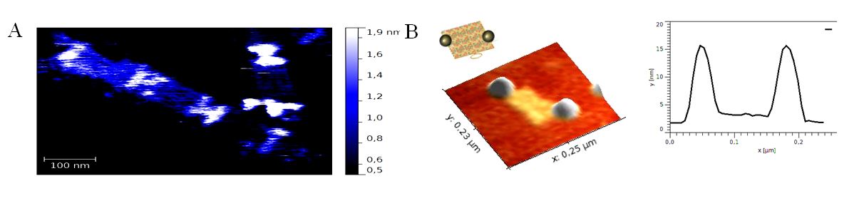
Dense metal nanostructure arrays with small interparticle distance allow for efficient focusing of incident light into a high density of nanoscale hotspots. The resulting high local electric fields can enhance optical processes at the metal surface. In principle, lithographic methods can be used to fabricate such nanostructured arrays, but the minimum achievable distance between patterned particles is typically limited in the lithographic process. Previous attempts to overcome this limitation require highly specialized fabrication approaches or particular structure designs [1].
We have developed a simple and general method to reduce interparticle distance in lithographically patterned arrays, involving repeated deposition and subsequent annealing of ultrathin gold films on top of pre-patterned gold nanoparticles. Surface diffusion of gold results in ripening of the initially patterned particles, thus making distance between particles smaller. The process can be tuned to realize a distinctly bimodal particle size distribution, suitable for focusing incoming radiation to a high density of nanoscale spots, with local field intensity enhancements up to three orders of magnitude compared to the incoming radiation, as confirmed by surface enhanced Raman scattering experiments and finite-difference time-domain calculations.
This work was supported by the Icelandic Research Fund, grant no. 110004022, the ESF network
Plasmon-Bionanosense, COST Action MP0803, and ERC starting grant no. 259432 (J.K).
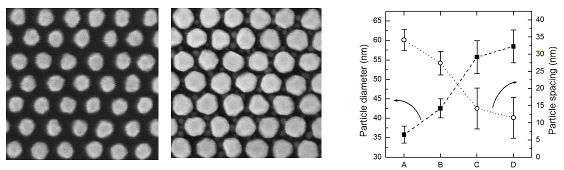
- Fig. 1: Gold nanoparticle array (lattice period 75 nm) patterned by e-beam lithography before (left image) and after (right image) additional deposition and annealing steps. The plot shows particle size and minimum spacing at different stages of the fabrication process.
[1] V. Merk, K. Leosson, J. Kneipp, Adv. Optical Mater. (2013) DOI: 10.1002/adom.201200061
1. Genetics and Cytology Department, Genetic Engineering and Biotechnology Division, National Research Centre, Dokki 12622, Cairo, Egypt
2. Cancer Biology Lab., Center of Excellence for Advance Sciences, National Research Centre, Dokki 12622, Cairo, Egypt
* Correspondence: Mona A. Abo-Zeid; e.mail: monaabozeid@yahoo.com
Background: Nanotechnology, an interdisciplinary research field involving chemistry, engineering, biology, and medicine, has great potential for early detection, accurate diagnosis, and personalized treatment of cancer. There is a new trend to use Gold nanoparticles (GNPs) with laser irradiation as a new photothermal therapy for cancer treatments.
Objective: The aim of the present studies was to evaluate the cytogenetic effects could be induced by high dose of rod shaped GNPs at 50nm diameter on normal female Swiss mouse bone marrow cells. This research work is a part from research project to evaluate the GNPs safe doses and rote of administration for Cancer Photothermal therapy.
Methods: The cytogenetic changes were evaluated by investigating the potential of GNPs to induce cytotoxicity and genetoxicity by injecting female Swiss mice i.v. with high dose of rod shaped GNPs at a concentration 37.5 ppm for 21 days (1 dose/week). The mice bone marrow cells were collected after 21 days to evaluate Chromosome Aberrations, Mitotic Index (MI), Sister Chromatid Exchanges (SCEs), Replicative Index (RI), Micronucleus in Polychromatic Erythrocytes (MNPCEs) and DNA damage by Comet assay.
Results: The data showed that GNPs at that high dose concentration induced chromosome aberrations and SCEs in mouse bone-marrow cells. The mean percentage of chromosome aberrations excluding gaps and SCEs/ cell increased in low significant manner in animal groups treated with GNPs in comparing to those of non-treated groups, however the animals treated with Cyclophosphamide (CP) as a positive control were found to have chromosome abnormalities and SCEs/ cell highly significantly in comparing to control. Also, GNPs induced micronuclei observed in bone-marrow PCEs in a mild significance manner. The MI and RI of the cells were decreased highly significantly (p<0.001), which were as high in significance as those induced by CP in comparing to non-treated groups. A high significant increase in comet tail length percentage and tail moment ratio indicating DNA damage were observed by Comet assay in animal groups treated with GNPs in comparing to non-treated groups. The mount of DNA damage in cells after treatment with GNPs was estimated from the comet tail length by the extent of migration of the genetic material.
Conclusion: The results indicated that rod shaped GNPs at high dose concentration and with repeated dose administration could induce DNA damage and reduce the cell proliferation highly significantly. However, the low doses of rod shaped GNPS could be safe to be used for Cancer Photothermal therapy (data not shown). Further research studies are required to optimize the safe doses of GNPs and rote of administration to be approved as new nanoparticles for Cancer Photothermal therapy by FDA.
Keywords: Gold Nanoparticles, Genotoxicity, Chromosome Aberrations, Sister Chromatid Exchanges, Micronucleus Test, Comet Assay
1 IPHT Jena, Albert Einstein Strasse 9, 07745 Jena, Germany
2 CEA Saclay, 91191 Gif/Yvette Cedex, France
3 CNRS, IRCELYON, Lyon, France
Nanomaterials have applications in various new fields like in the field of environmental catalysis (or photocatalysis), i.e. new processes for green chemistry. Indeed, the high dispersion of the active phase (oxide, metal) strongly increases the catalytic efficiency. In order to get such nanomaterials, laser pyrolysis is a promising method for the synthesis of various nanoparticles, with well defined chemical composition, size and structure and allows the use of gaseous or liquid precursors [1, 2]. This gas phase synthesis method is based on the interaction of metal-organic precursor droplets in aerosol with a strong CO2-laser with appearance of a flame where nanopowders nucleate and grow. Aim was the preparation of Au-TiO2 hybrid nanoparticles. TiO2 and noble metal nanoparticles are known as (photo)catalytic materials. Au nanoparticles show plasmonic properties and are used as markers in bioanalytics.
In order to synthesize the Titanium oxide, TTIP (Titanium Tetra isopropoxide) was chosen as Ti precursor. Gold was introduced as HAuCl4 in an ethanol solution or as wet chemically precipitated gold nanoparticles with a content of up to ≈1 wt% in the final powder. The particles were characterized structurally and optically.
We also compared the photocatalytic activity of TiO2 (commercial product P25 and laser pyrolyzed particles) and Au-TiO2 hybrid particles by evaluation of the degradation of formic acid under UV illumination.
Our work was supported by DLR/BMBF, project AQUATEST 01DQ12090.
[1] W.R. Cannon, S.C. Danforth, J.S. Haggerty and R.A. Marra, J. Am. Ceram. Soc, 65, 330 (1986)
[2] M. Cauchetier, O. Croix, N. Herlin, M. Luce, J. Am. Ceram. Soc. 1994, 77(4), 993-8
1 Institute for Life sciences, Physics and Astronomy, University of Southampton, Highfield, SO17 1BJ, Southampton, UK
2 Institute for Life sciences, Faculty of Medicine, University of Southampton, Highfield, SO17 1BJ, Southampton, UK
The potential of small metal nanoparticles for converting resonant light into local heat has given rise to new applications in biomedicine [1]. While many studies focus on destructive laser hyperthermia of malignant cancer cells, the impact of photothermal therapies using plasmonic nanoparticles on healthy, primary human cells is of interest for controlling biological functions and for combined, dual-action treatments. Here, we present results on the controlled laser treatment of human endothelial cells (HUVECs) using oligo-ethylene glycol (OEG-)coated nanoparticles, which are either specifically targeted using peptide-functionalization, or which are nonspecifically taken up by the cells without further functionalization [2-4]. Peptide-functionalized nanoparticles were found to specifically bind to VEGF-receptors in the cell membrane [2]. Subsequent laser treatment at mild conditions of up to 30 W/cm2 laser intensity resulted in partially reversible damage of the cellular membrane. A remarkable recovery of cells was found within 24 hours following treatment [3]. For non-functionalized nanoparticles, combined ICP-OES analysis and numerical modeling indicated that irreversible cell apoptosis was caused by collective heating effects over the illumination spot (Fig. 1) [3]. It is argued that the multiscale effects of nanoparticle hyperthermia range from single nanoparticles to collective heating of macroscopic areas, and on time scales from picoseconds to seconds, are an important factor in designing effective nanoparticle treatments. New studies explore plasmonic laser treatment on in-vitro angiogenesis, the organisation of endothelial cells into a network of microvessels in a matrigel (Fig. 2).
- Fig. 1: Calculated temperature increase caused
by a single nanoparticle (left) and integrated
over the 500m illumination spot (right) [4].
Fig. 2: In-vitro angiogenesis of HUVECs in a MDA cancer-cell medium.
[1] Dreaden, E. C., Alkilany, A. M., Huang, X., Murphy, C. J., and El-Sayed, M. A., Chem. Soc. Rev., 41, 2740-2779 (2012); Barreto, J. A., O'Malley, W., Kubeil, M., Graham, B., Stephan, H., Spiccia, L., Adv. Mater., 23, H18-H40 (2011).
[2] Bartczak D, Sanchez-Elsner T., Louafi F., Millar T., Kanaras A. G., Small 7, 388–394 (2011).
[3] D. Bartczak, O. L. Muskens, T. M. Millar, T. Sanchez-Elsner, A. G. Kanaras, Nano Lett. 11, 1358-1363 (2011).
[4] D. Bartczak, O. L. Muskens, S. Nitti, T. Sanchez-Elsner, T. M. Millar, and A. G. Kanaras, Small 8, 122-130 (2012).
Leibniz-Institut für Polymerforschung Dresden e.V., Hohe Straße 6, 01069 Dresden, Germany.
Nanoparticles have to be placed at certain sites for most practical applications. A very efficient approach for mass production using thermoplastics is injection moulding. However, the processes of melt flow in nanometre dimensions are not yet quite clear. Therefore, we investigate the immobilisation of nanoparticles in the surface layer of polycarbonate during moulding [1]. AuNP were assembled on a substrate using electrostatic adsorption from solution. A cationic polymer (PEI) was deposited in advance. This process was repeated several time to increase the degree of coverage. The substrate was then placed in the mould of an injection moulding machine, and the melt was injected. We investigated the arrangement of the AuNP in the surface layer of the melt. The TEM micrograph in figure 1 shows, that, surprisingly, the part surface is very even in the range of some nanometres. The AuNP were surrounded by PEI. PEI shows the typical reactions of a polyamine, e.g. complex formation with Cu2+, as EDX measurements revealed. The AuNP are still catalytically active. That is why both compounds, the AuNP and PEI are still accessible for small molecules of a solution, brought in contact with the part surface. We found out, that the embedding depth depend on the thickness of the PEI layer. This is used in further work to gain control over the arrangement of nanoparticles on the part surface. The approach could be the basis for the production of components used in material's engineering, in which metal nanoparticles transfer light into heat. The components could be used in electronics, e.g. sensors, switches, and memory devices.
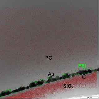
- Fig. 1: EFTEM image of AuNP embedded at a polycarbonate surface layer. The AuNP are still surrounded by the functional polymer PEI. C and SiO2 were deposited by PVD after moulding.
[1] 38. Jürgen Nagel, Petchara Chunsod, Cordelia Zimmerer, Frank Simon, Andreas Janke, Gert Heinrich, "Immobilization of Gold Nanoparticles on a Polycarbonate Surface Layer During Molding", Mater. Chem. Phys. 129 (2011) 599– 604
MoNOS, Leiden Institute of Physics, Leiden University
Postbus 9504, Niels Bohrweg 2, 2300 RA Leiden, Netherlands
Compared to electron or scanning probe microscopies, optical studies of single objects in far field is non-invasive, reaches beyond surfaces, and opens many time-resolved and frequency-resolved experiments. Unique insights are provided by these signals into the dynamics of nano-objects and of their surroundings [1].
i) We study single gold nanoparticles by photothermal and pump-probe microscopy and detect their acoustic oscillations launched by a pump pulse [2]. These experiments can be done in an optical trap, where a single nanorod orients along the polarization of the trapping laser. We could thus clarify the origin of the damping of mechanical vibrations in gold nanoparticles.
ii) Photothermal microscopy opens the study of non-fluorescent absorbers, down to single-molecule sensitivity [3]. Combining photothermal contrast with photoluminescence, we can measure the luminescence quantum yield on a single-particle basis and gain insight into the complex relaxation phenomena leading to emission by metal particles. Moreover, the high signal-to-noise ratio of this contrast mechanism opens up uses of individual gold nanoparticles for local plasmonic and chemical probing. Binding and unbinding events of single protein molecules can be detected in this way [4].
iii) Gold nanorods generate strong field enhancements near their tips. By matching the rods' aspect ratio to a dye's fluorescence and excitation spectra, we could observe enhancements in excess of thousand-fold for the fluorescence of single Crystal Violet molecules [5]. Such high enhancements have been observed before with bowtie nanoantennas. We show that gold nanorods can produce equally high local fields thanks to their narrow plasmon resonance, but they are much easier to synthesize, functionalize and disperse in solution than lithographically-made nanostructures.
- Fig. 1: Example of an absorption trace of a single gold nanorod showing binding and unbinding events of single proteins [4].
[1] F. Kulzer et al., Angew. Chem. 49 (2010) 854.
[2] A. L. Tchebotareva et al., Laser Photon. Rev. 4 (2010) 581-597.
[3] A. Gaiduk et al. Science 330 (2010) 353
[4] P. Zijlstra et al., Nature Nanotech. 7 (2012) 379.
[5]. HF. Yuan et al., Angew. Chem. 51 (2013) .
Institute of Photonic Technology, Albert-Einstein-Str. 9, 07745 Jena, Germany
Cylindrical nanophotonic devices in fiber form are a new class of integrated components which can overcome several limitations of state-of-the-art planar devices such as insufficiently small aspect ratios or significant insertion loss when coupled to single-mode fibers. Our approach to overcome these essential bottlenecks and to realize such functionalized fiber waveguide structures is based on pressure-assisted melt-filling of holey photonic crystal fibers (PCFs) [1, 2]. This method gives PCF new functionalities and opens up new ways for applications of optical fiber.
The structures I will discuss here are modified PCFs including metallic nanowire (Fig. 1a), giving rise to so-called plasmonic PCFs [3].
I will discuss the basics of plasmonic excitation on metallic nanowires and show that the propagating plasmons can be described as planar plasmons spiraling around the wire circumference. Also I will introduce ultralong range plasmons (Fig. 1b), which reveal much longer propagation lengths than their planar counterparts. After that I will focus the talk onto several experimental implementations of plasmonic fiber by showing plasmonic molecules, local near field polarizations of plasmonics supermodes and broadband azimuthal polarizers.
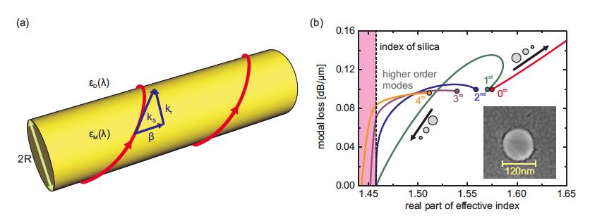
- Fig. 1: (a) Schematic of a spiralling surface plasmon mode on a single isolated metal wire (red lines represent plasmon trajectory on the wire surface). Dark blue arrows indicate the wave vector diagram of the propagating plasmon modes (ks: planar surface plasmon mode wave vector, β: longitudinal propagation constant, kt: quantized tangential wave vector). (b) Complex plane representation of the effective mode index of the different plasmon modes as function of wire radius (silver wire). Each curve starts at a radius of 800nm (coloured points) and follows the complex effective mode index for a reducing wire radius (radius reduction indicated by the black arrows and grey circles, wavelength for all calculation: 600nm). The numbers stand for the mode order of the indicated plasmon mode (order of Bessel function). In the light red region the real part of the plasmon modes falls below the index of silica (nsilica(600nm) = 1.458). The inset depicts the smallest wire (120 nm diameter) fabricated so far with the pressure-assisted melt infiltration technique.
[1] M. A. Schmidt et al., Phys. Rev. B. 77 (2008), 033417.
[2] P. Uebel et al., Opt. Exp. 20 (2012, 28409.
Institute for Physical and Theoretical Chemistry, Braunschweig University of Technology, 38106 Braunschweig, Germany
E mail: t.sen@tu-bs.de
Over the past years, the interaction of fluorescent dye molecules with plasmonic nanostructures has been studied extensively both theoretically and experimentally [1-3]. Metallic nanoparticles can influence the electronic transitions of a luminescent molecule in the absorption and the emission channel. Metallic nanoparticles are also capable of influencing the Förster resonance energy transfer (FRET) between two close molecules. FRET is widely used as a spectroscopic tool in lighting, single-molecule spectroscopy, imaging, and sensing [4-6]. Here, we discuss on single-molecule FRET between a donor-acceptor pair near Gold nanostructures. Accurate positioning and alignment of nanoparticle and fluorophore with respect to each other and the incoming electric field was achieved by a three-dimensional, self-assembled DNA origami. A three-dimensional, pillar-shaped DNA origami was folded from one M13mp18- derived scaffold strand and 207 staple strands (Fig. 1) [7]. During hybridization, two dye-labeled strands (ATTO647N and Cy3B) and three to six capturing strands were additionally incorporated at the half height of the pillar. The distance between the donor-acceptor pair is 6 nm. DNA functionalized Au NPs were bound to the pillar by hybridization to the capturing strands. We designed a gap of 23 nm between the NPs as a compromise of strong fluorescence enhancement. We have studied the efficiency and rate of FRET between single dyes in the vicinity of gold NP monomers and dimers of varying sizes. We also include a discussion about the fluorescence enhancement by Au core Ag shell nanoparticles of different size using similar origami structure. TEM images of as prepared Au core Ag shell nanoparticles revealed the average sizes of 24, 56 and 108 nm. These bimetallic core-shell nanoparticles were functionalized with thiolated single-stranded DNA and were bound to the pillar origami by hybridization to the capturing strands.
Funding by ERC is gratefully acknowledged.
- Fig. 1: Sketch of the DNA origami with two AuNPs and fluorescent dye at the equator plane.
[1] L. Novotny and N. van Hulst, Nat Photon 5 (2011) 83-90.
[2] F. Reil, U. Hohenester, J. R. Krenn and A. Leitner, Nano Lett. 8 (2008) 4128-4133.
[3] G. P. Acuna et al., ACS Nano 6 (2012) 3189-95.
[4] T. Förster, Ann. Phys. 437 (1948) 55.
[5] J. Zhang, Y. Fu and J. R. Lakowicz, J. Phys. Chem. C 111 (2007) 50-56.
[6] V. Faessler, C. Hrelescu, A. A. Lutich, L. Osinkina, S. Mayilo , F. Jäckel and J. Feldmann Chem. Phys. Lett. 508 (2011) 67–70.
[7] G. P. Acuna, F. M. Moller, P. Holzmeister, S. Beater, B. Lalkens and P. Tinnefeld, Science 338 (2012) 506-10.
Institute of Photonic Technology, Albert-Einstein-Str. 9, Jena, Germany
Institute of Physical Chemistry and Abbe Center of Photonics, Helmholtzweg 4, Jena, Germany
* Department of Physics, University of Osnabrück, Barbarastraße 7, Osnabrück, Germany
The application of metal-molecule interfaces bares a huge potential for applications in various fields of chemistry as the properties and functions of metal nanoparticles can be modified and extended by surface-functionalization. The work presented aims at the functionalization of complex colloidal particles, in particular Au nanopeanuts and Au/Pt/Au nanoraspberries (Fig. 1) fabricated by the galvanic replacement method with photoactive molecular electron donors.[1] As functionalizing agent the commercially available Ru-complexes N719, N749 and Z907 are used, which are commonly applied as photosensitizers in dye-sensitized solar cells.[2] A detailed investigation of the conjugates by means of Raman spectroscopy not only proves the successful surface-functionalization but also reveals the geometrical and electronic structure of the adsorbed complexes. It is shown that the electronic structure of the ruthenium complexes is retained upon binding to the metal surface.[3] A detailed investigation of the SERRS signal dispersion and the enhancement profile is performed by applying micro-fluidics.[1] This technique allows for an efficient and reproducible wavelength screening of the SERRS signal in colloidal solutions. The enhancement profiles obtained differ from the extinction spectra of the nanoparticles, indicating that electronically resonant processes are involved in the Raman signal enhancement of the investigated nanoparticle conjugates in addition to the Raman signal enhancement observed due to the SERS effect.
We gratefully acknowledge funding of the research project 'Photonic Nanomaterials' - PhoNa (Grant No. 03IS210EA) within the framework 'Spitzenforschung und Innovation in den Neuen Ländern'.
- Fig. 1: TEM image of the Au nanopeanuts (a) Au/Pt/Au nanoraspberry (b) and schematic illustration of the Ru-dye functionalization (c).
[1] Theil, F.; Zedler, L.; März, A.; Xie, W.; Csáki, A.; Fritzsche, W.; Cialla, D.; Schmitt, M.; Popp, J.; Dietzek, B., JPCC 117, 1121-1129 (2013).
[2] Listorti, A.; O'Regan, B.; Durrant, J. R., Chem. Mater. 23, 3381–3399 (2011).
[3] Zedler, L.; Theil, F.; Csáki, A.; Fritzsche, W.; Rau, S.; Schmitt, M.; Popp, J.; Dietzek, B. RSC Advances 2, 4463-4471 (2012).
*Institute for Photonic Technologies, Dept. of NanoBiophotonics, Albert-Einstein-Str. 9, 07745 Jena, Germany
**Ilmenau University of Technology, Institute for Chemistry and Biotechnology, Dept. of Physical Chemistry and Micro Reaction Technology, Weimarer Str. 32, D-98693 Ilmenau, Germany
Noble metal nanoparticles are well known for their plasmonic activity, which plays an important role in particle based analysis [1]. Since shape anisotropic nanoparticles have - in contrast to spherical ones - a sharper and over the whole visible spectra tunable plasmonic peak [2], the synthesis of such well-defined particle structures is preferred.
The preparation of anisotropic nanoparticles can be divided into two parts: a) the synthesis of seed particles and b) their subsequently growing [3]. The critical part belongs to the synthesis part and has to be well controlled. To improve this controlling, we will present a method for the synthesis of silver seed nanoparticles by use of microfluidic devices. With a microfluidic system consisting of three different micro mixers we will show the need of special adopted mixers for the efficient synthesis and their influence on seed particle formation. Each mixer includes a certain mixing regime to decrease the diffusion distances, which results in fast mixing of the reagents as an essential starting for seed production and gives highly reproducible results. Furthermore will be shown, that this microfluidic synthesis results in smaller, less disperse and more sensitive silver seed particles compared to the ones made via batch synthesis in a flask. Another advantage of the microfluidic synthesized seeds is the increased ability to grow prisms in defined sizes with higher quality and quantity. The microfluidic system represents an approach for higher throughput and enables so a controllable and continuous synthesis of nanoparticles as a fundament for an automated system for large scale nanoparticle generation.
[1] Mayer K. M. and Hafner J. H., (2011), Localized surface plasmon resonance sensors, Chemical reviews, Vol.111 No.6, 3828-3857
[2] Niu W., Zhang L. and Xu G., (2013), Seed-mediated growth of noble metal nanocrystals: crystal growth and shape control, Nanoscale,
[3] Aherne D., Ledwith D. M., Gara M., et al., (2008), Optical Properties and Growth Aspects of Silver Nanoprisms Produced by a Highly Reproducible and Rapid Synthesis at Room Temperature, Advanced Functional Materials, Vol.18 No.14, 2005-2016
Nanoscience Center, Department of Physics, P.O. Box 35, 40014 University of Jyväskylä, Finland
* Institut für Physik, Karl-Franzens-Universitt Graz, Universittsplatz 5, 8010 Graz, Austria
** School of Engineering and Applied Sciences, Harvard University, Cambridge, MA 02138, USA
During the last decades surface plasmons polaritons (SPPs) have attracted vast interest because of their strongly enhanced light-matter interactions, which opens routes towards optical devices beyond the diffraction limit allowing nanoscale integration of photonics. Of particular interest is also strong coupling between SPPs and molecular excitations (ME), which manifests itself through the formation of new hybrid modes exhibiting Rabi splittings even up to several hundreds of meV depending on the molecular species and concentration [1]. The obtained new hybrid modes, i.e. plexitons, could provide the non-linearity to enable interactions between photons/plasmons, needed for active all optical components.
We have studied strong coupling between SPPs and various molecules [2], and shown that in all of them the fluorescence of the MEs reveals the coexistence of strongly coupled plexitons and non-coupled MEs. As the molecular fluorescence is directly related to the ME occupation, it provides an ideal tool for studying the polariton dynamics. Here we have utilized dye molecules with an especially large Stokes shift, i.e. Nile Red (NR), to demonstrate the absence of scatterings among the plexitons and to show that the modes decay via dephasing and internal relaxation of the molecules to a fluorescing state of the dye [3]. This result provides essential information on the dynamics of the strongly coupled modes.
- Fig. 1: a: Scattering spectra measured from a NR-sample at different angles and polarizations: TE (red) and TM (blue). The TE spectra match well with the fluorescence of NR, while TM shows the scattered plexitons. b-c: Measured total fluorescence (circles) for three samples together with the transfer matrix calculations (solid lines in b) and the quantum mechanical three state model (solid lines in c).
[1] A.M. Glass, et al. Opt. Lett. 5 (1980) 368; J. Bellessa, et al, Phys. Rev. Lett. 9 (2004) 036404; J. Dintinger, et al. Phys. Rev. B 71 (2005) 035424; T.K. Hakala, et al. Phys. Rev. Lett. 103 (2009) 053602; P. Vasa, et al. Nature Phot. 7 (2013) 128.
[2] S.V. Baieva, et al. Nanoscale Research Letters 7 (2012) 191; S.V. Baieva, et al. J. Chem. Phys. 138 (2013) 044707.
[3] M.A. Koponen, U. Hohenester, T.K. Hakala, and J.J. Toppari, submitted (2013).
Institute for Photonic Technologies, Dept. of NanoBiophotonics, Albert-Einstein-Str. 9, 07745
Jena, Germany
Silver nanoprisms are promising transducers for bioanalytical applications due to their strong enhanced localized plasmonic fields in comparison to other nanoparticles [1]. These applications often require immobilized particles on substrates in environmental condition. But silver is more vulnerable to oxidation in atmospheric condition as gold [2].
Therefore the objective of this work is the investigation of aging effects of immobilized silver nanoprisms due to their material property. The behavior of aging effects will be investigated dependent on different surrounding environments and substrate modification.
Morphological transformations were characterized by long-term Scanning Force Microscopy (SFM) measurements. These transformations start at nanoprisms corners and edges within one day. The material transformation also leads to plasmonic changes, which were investigated by dark-field single particle spectroscopy measurements. These investigations shown a red shift and decreased dipole resonance within three hours.
Furthermore an Atomic Layer Deposition (ALD) passivation was applied to prevent these changing effects. The used passivated material was an aluminum oxide layer, which was applied by a 30°C oxygen plasma enhanced ALD.
- Fig. 1: a: Dark-field single particle spectrum of a immobilized silver nanoprism over a period of eight hours.
b: Scanning force microscopy time-series over a period of five hours.
[1] Matthew Rycenga, Claire M. Cobley, Jie Zeng, Weiyang Li, Christine H. Moran, Qiang Zhang, Dong Qin, and Younan Xia, Chem. Rev.111 (2011)
[2] Kathryn M. Mayer†, Jason H. Hafn, Chem. Rev. 111 (2011)
University of Duisburg-Essen, Universitaetsstr. 5, D-45141 Essen, Germany
Composite plasmonic nanoassemblies exhibit fascinating optical properties. One interesting application is their use in surface enhanced Raman scattering (SERS) [1, 2]. However, the chemical synthesis of plasmonic nanostructures with well defined geometries is still a challenge. In this contribution, anisotropic nanoassemblies, composed of gold nanorods (GNRs) as the core and gold nanoparticles (GNPs) as satellites, were synthesized (see Fig.1). Typical TEM images (Fig. 2A) of the nanoassemblies demonstrate their well-defined geometry. The position of the tunable localized surface plasmon resonance (LSPR) peak can be controlled by the aspect ratio of the core (GNR) and the size of the satellites (GNPs). Due to plasmonic coupling and the resulting hot spots in the particle junctions (FEM calculations, not shown), the nanoassemblies are excellent SERS substrates. Finally, we used the nanoassemblies for the detection of the cytokine interleukin-6 in a SERS immunoassay. IL-6 could be detected at concentrations as low as 0.1pg/mL with great specificity (Fig. 2B).
Funding by the Alexander von Humboldt foundation (AvH) and the Deutsche Forschungsgemeinschaft (DFG) is gratefully acknowledged.
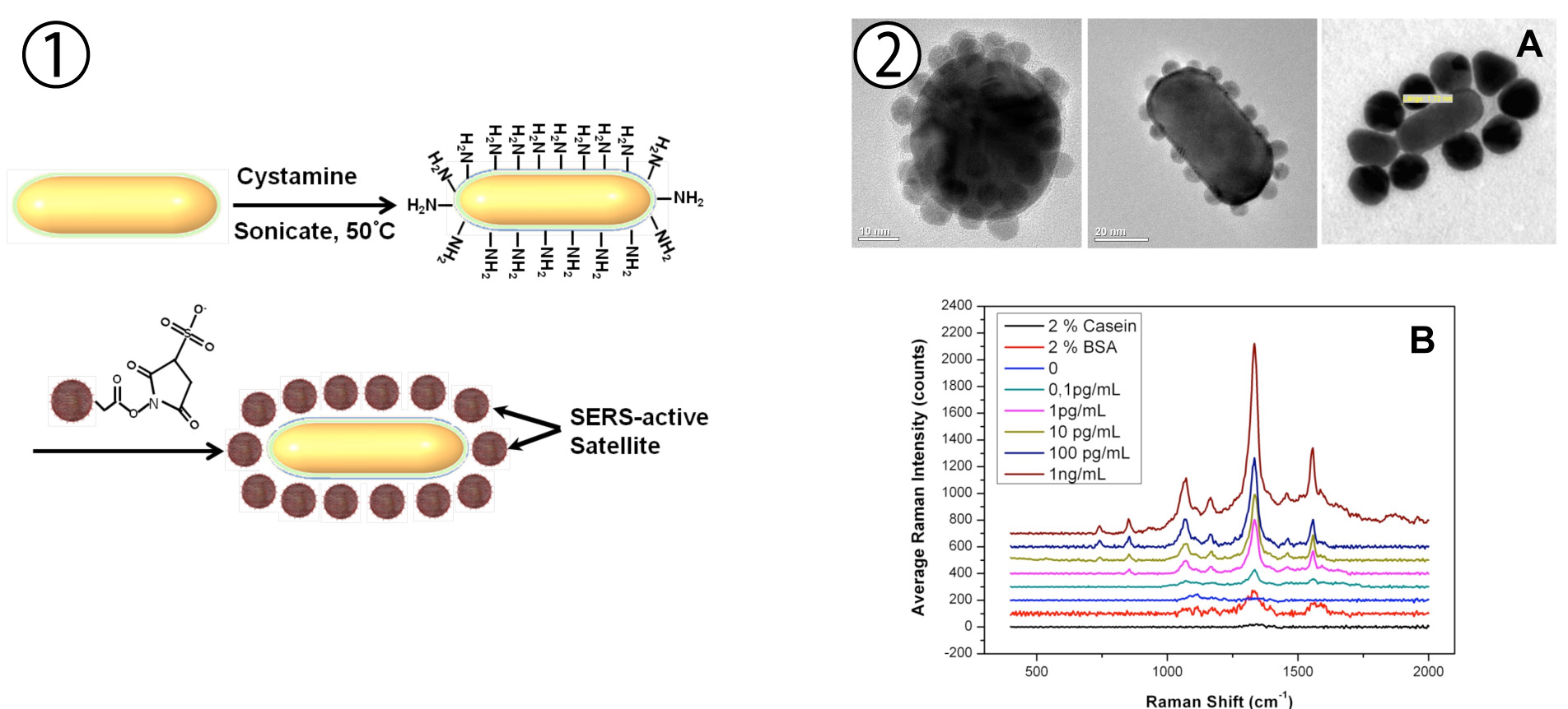
- Fig. 1: Scheme for the synthesis of anisotropic plasmonic core (GNR)/satellite (GNPs) nanoassemblies
Fig. 2: a: (A) TEM images of anisotropic plasmonic nanoassemblies with various aspect ratios of GNRs and sizes of satellite GNPs (5 nm and 20 nm), respectively. (B) Their use for the SERS detection of IL-6 in an immunoassay.
[1] L. Vigderman, Adv.Mat. 24 (2012) 4811-4841.
[2] R. A. Alvarez-Puebla and L.M.Liz-Marzan, Chem. Soc. Rev.41 (2012) 43-51.
1 SATIE (CNRS UMR8029), Ecole Normale Supérieure de Cachan-Bretagne, Campus de Ker Lann, F-35170 Bruz, France
2 Laboratoire Aimé-Cotton (CNRS UPR3312), Université Paris Sud, Bât. 505, Orsay, France
Isolated gold nanoparticles in liquid suspension can effectively quench the fluorescence of molecules that are nearby (< 10 nm), e.g. fluorophores attached to the particles via molecular linkers. Using fluorescence correlation spectroscopy, which analyses the fluorescence from individual objects in a small probe volume, we have been able to demonstrate unambiguously the quenching of ligated chromophores, and to identify spontaneously desorbed ligands that display fluorescence [1,2].
Enhancement of molecular spectroscopic responses may occur at 'hot spots' in aggregates of plasmonic nanoparticles. Moreover, such aggregates display an intense, and specific, resonant light scattering, which in itself provides a useful signal for optical detection at low concentrations in small volumes [3]. We are currently investigating the reversible aggregation of various functionalised gold nanoparticles, using microfluidic systems [4] as a platform for precise control of the aggregation as well as for the development of new (molecular plasmonic) biosensing schemes.
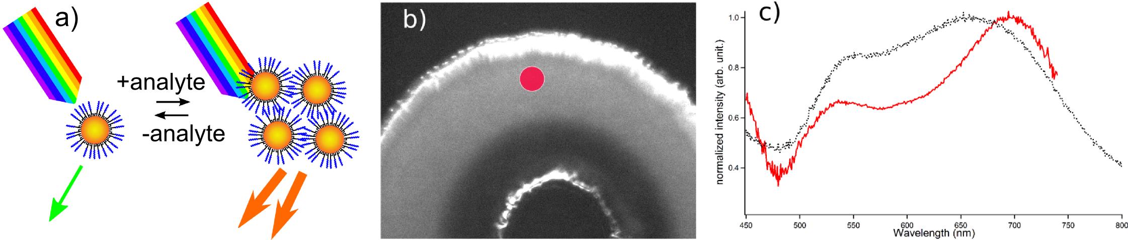
- Fig. 1: (a) Reversible aggregation of functionalised gold colloids in the presence of an analyte leading to a change in resonant light scattering. (b) Light scattering by gold colloid aggregates in a microfluidic channel under dark-field illumination. (c) Microspectroscopy (red curve) showing the NIR plasmonic scattering resonance of the aggregates. The black line is the resonant light scattering from a bulk suspension of aggregated particles.
[1] M. Loumaigne, R. Praho, D. Nutarelli, M. H. V Werts and A. Débarre, Phys. Chem. Chem. Phys. 12 (2010) 11004–11014
[2] J. R. G. Navarro, M. Plugge, M. Loumaigne, A. Sanchez-Gonzalez, B. Mennucci, A. Débarre, A. M. Brouwer, M. H. V Werts, Photochem. Photobiol. Sci. 9 (2010) 1042–1054
[3] J. R. G. Navarro and M. H. V Werts. Analyst 138 (2013) 583–592
[4] M. H. V. Werts, V. Raimbault, R. Texier-Picard, R. Poizat, O. Français, L. Griscom and J. R. G. Navarro,
Lab on a Chip 12 (2012) 808–820
Institute of Chemistry, The Hebrew University of Jerusalem, Jerusalem 91904, Israel
DNA machines represent supramolecular organized nanostructures that perform fueled (or anti-fueled) reversible mechanical transitions. Our laboratory has developed different DNA machines, including tweezers, walkers, catenated rings, and more. Recently, we applied DNA machines as functional devices for the cyclic and reversible fuel-driven organization of programmed Au nanoparticle nanostructures, or fluorophore/Au NPs nanostructures. Interparticle plasmonic coupling phenomena in the Au NPs nanostructures are observed, and surface- enhanced fluorescence/fluorescence quenching processes are programmed by the fluorophore/Au NPs nanostructures generated by the DNA machines. This will be exemplified by the following systems:
1. A three-ring catenane system is used for the dynamic programming of Au NPs nanostructures. The effect of the nanoparticle structures on the plasmonic features of the systems will be described, and the experimental results will be theoretically modeled. Effects of NPs sizes on the plasmonic properties of the systems will be discussed.
2. The decoration of the three-ring catenane system with Au NPs and a fluorophore lead to dynamically dictated nanostructures, revealing surface- enhanced fluorescence or quenching phenomena. The experimental results are theoretically modeled and correlated with the structural properties of the systems.
3. "Tweezers" DNA machines are used as molecular devices that control plasmonic properties of fluorophores. The tailoring of Au NP/fluorophore-functionalized tweezers leads to surface-enhanced fluorescence or fluorescence quenching phenomena upon the reversible opening and closure of the "tweezers".
4. The photophysical properties of other Au NPs-functionalized DNA machines, such as a structurally programmed Au NPs dumbbell DNA rotaxanes or switchable fluorophore/Au NPs DNA muscles, will be discussed.
1 Institute of Photonic Technology, Albert-Einstein-Straße 9, 07745 Jena, Germany 2 Nanoscience Center, Department of Physics, P.O. Box 35, FI-40014 University of Jyväskylä, Finland
The interaction between metal nanoparticles and light offers a high potential in the development of new photonic devices for biomedical applications. Thereby, the nanoparticle-light-interaction can be used either for biosensor-applications with a sensitive detection of analyte molecules or for a contact-free manipulation of biological matrices immediately adjacent to a highly excited nanoparticle.
In our studies we're investigating the effects of highly excited metal nanoparticles on stretched DNA molecules, driven by femtosecond laser pulses. Our experimental setup consists of silver nanoparticle (AgNP) labeled DNA molecules immobilized on a radiation sensitive Polymethylmethacrylate (PMMA) layer in air. By the exposure of selected AgNP-labeled DNA molecules with fs laser pulses we determined the effects on the DNA molecules by the destruction of the underlying PMMA surface. Thereby, we observed an energy coupling into the DNA molecules leading to a guiding of this energy along the molecular structure resulting in a destruction of the PMMA layer reaching a distance up to 4 μm (Fig.1). We suppose that this guiding is caused by the escape of electrons of the excited nanoparticle [1]. To underline our hypothesis we've repeated the experiment with fluorescence-labeled DNA molecules, resulting in bleaching of the DNA over similar distances (Fig. 2).
This novel excitation transfer phenomenon in molecular wires could have implications as interconnects and functional devices in molecular electronics as well as for biosensing applications [2].
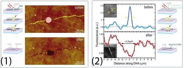
- Fig. (1): Scheme with AFM images of AgNP-labeled DNA molecules before and after their treatment with fs laser pulses.
Fig. (2): Scheme and fluorescence images of AgNP-labeled DNA molecules before and after their treatment with fs laser pulses.
[1] Wirth, J., et al., Plasmonic Nanofabrication by Long-Range Excitation Transfer via DNA Nanowire. Nano Letters, 2011. 11(4): p. 1505-1511.
[2] Toppari, J.J., et al., Plasmonic Coupling and Long-Range Transfer of an Excitation along a DNA Nanowire. Acs Nano, 2013. 7(2): p. 1291-1298.
Department of Chemistry, University of Duisburg-Essen, 45141 Essen, Germany
Monitoring of heterogeneous catalysis requires surface-selective analytical methods because the chemical transformations are confined to the solution-catalyst interface. Surface-enhanced Raman scattering (SERS) is a surface-selective technique with high sensitivity and chemical specificity which fulfills the requirements for in situ monitoring of molecular transformations in heterogeneous catalysis. However, catalytically active materials such as Pt, Pd, and small Au nanoparticles (NPs) do not provide sufficient plasmonic activity for the required SERS enhancement. Bifunctional nanostructures with both plasmonic and catalytic activity can enhance the Raman signal of chemical species involved in the catalytic reactions. Au-Pt-Au core-shell nanoraspberries with a high Pt surface area and a high plasmonic activity represent an example of bifunctional hybrid NPs for combined catalysis/SERS monitoring.[1] Chemical reactions catalyzed by Pt can be monitored by SERS due to the plasmonic Au protuberances grown on the Pt surface. Alternatively, the catalysts NPs can be self-assembled on a large plasmonically active core to form a plasmonic superstructure; in this case the chemical species on the catalyst surface experiences a sufficient SERS enhancement (Fig.1).[2] In a proof-of-concept study, rationally designed bifunctional Au NP superstructures were employed for direct and label-free monitoring of the metal NP-catalyzed reduction of 4-nitrothiophenol (4-NTP) to the corresponding aniline derivative (4-aminothiophenol, 4-ATP).
Support by the Alexander-von-Humboldt Foundation and the German Research Foundation is gratefully acknowledged.
- Fig. 1: SERS monitoring of heterogeneous catalysis by using 3D plasmonic Au superstructures.
[1] W. Xie, C. Herrmann, K. Kömpe, M. Haase and S. Schlücker, J. Am. Chem. Soc. 133 (2011) 19302-19305.
[2] W. Xie, B. Walkenfort, and S. Schlücker, J. Am. Chem. Soc. 135 (2013) 1657-1660.
National Institute of Laser Enhanced Sciences, Cairo Univ, 12613 Giza-Egypt.
Photodynamic therapy (PDT) is an established anticancer treatment employing a photosensitizer, visible light and oxygen. In both photodynamic therapy (PDT) and photodynamic diagnosis (PDD) of tumors, the photosensitizer is preferentially taken up by the malignant cells. The photosensitizer alone is harmeless either to normal or diseased cells, then illumination with light of the appropriate wavelength and dose can induce photochemical reactions resulting in tissue destruction via type I/II mechanism and/or emitting fluorescence which can be used for several biological applications. Nanoparticles (NPs) and plasmonics are increasingly finding wide applications in many areas related to biomedical diagnostics and therapeutics due to their unique electromagnetic, physical and chemical properties. The presence of metallic surfaces or particles in the vicinity of a fluorophore can dramatically alter the physicochemical properties of a fluorophore. Here we offer an attempt to enhance the fluorescence emissions of two powerful photosensitizing agents, hypericin and rose bengal, by conducting AgNPs (10-12 nm in size). This study can provide a high contribution for the photodynamic dyes in photodetection and photodiagnosis of tumors.
Fachbereich Physik und Wissenschaftliches Zentrum für Materialwissenschaften (WZMW), Philipps Universität Marburg, Germany
Early reports indicated that the engineered polyelectrolyte multilayer (PEM) microcapsules (diameter from 1 to 10µm) can be applied for targeted delivery in tumoral cells. Even though several strategies to load these capsules exist, they are still limited for cargo molecules with reduced solubility in water like most of the anti-cancer drugs. The encapsulation of micelles in such capsules and the further encapsulation the cargo molecule via hydrophobic interactions offer the possibility to encapsulate a broad number of molecules with biological interest. This method developed by Tong et al [1]. is based in the co-precipitation of polystyrene-block-poly( acrylic acid) (PS-b-PAA) micelles with CaCO3 that acts as a template in the formation Layer-by-Layer (LbL) of the PEM capsules. In our work, we will show that it was possible to encapsulate the one hydrophobic substrate of the phosphatase enzyme. We will show that it was possible to release this hydrophobic cargo in the cytoplasm of cancer cells through light-triggered opening of the capsule and we were able to observe by fluorescence microscopy the enzymatic reaction between the phosphatase enzyme and its substrate in the cellular cytoplasm[2]. Our work, shows that it is possible to remotely trigger biological reactions inside cells. Light-responsive capsules were used for this purpose.
[1] W. Tong et al."Micelles-encapsulated microcapsules for sequential loading of hydrophobic and water-soluble drugs" Macromol Rapid Commun. 31, 1015-1019 (2010).
[2] M. Ochs. S. Carregal-Romero et al "Light-addressable capsules as caged compound matrix for controlled in vitro release" Angewandte Chemie International Edition. 52, 695-699 (2013).
1 Institute of Physical Chemistry and Abbe- Center of Photonics, Friedrich-Schiller-University Jena, Helmholtzweg 4, 07743 Jena, Germany 2 IPHT - Institute of Photonic Technology, Albert-Einstein-Straße 9, 07745 Jena, Germany
Surface enhanced Raman spectroscopy (SERS) is an attractive tool in chemical and biological sciences because of not only its fingerprint specificity but also its high sensitivity [1]. SERS effect is based on the strong plasmonic field enhancement at rough metallic surfaces [2]. For this purpose, an easy fabrication technique of silver SERS substrates is established in this work. Enzymatically generated silver-nanoparticles (EGNPs) [3, 4] are prepared and coated with different layer thicknesses of SiO2 applying atomic layer deposition (ALD). The growth of 3D flower-like nanostructures is observable after the deposition of 10nm SiO2. Due to the rough surface, an efficient and reproducible SERS active substrate is generated. In this study, the morphological studies as well as the SERS response of these 3D nanostructures in combination with possible quantitative analysis are introduced.
Funding the projects "QuantiSERS"and "Janaer Biochip 2,0" within the framework "InnoProfile Transfer- Unternehmen Region" the
Federal Ministry of Education and Research, Germany (BMBF) is gratefully acknowledged.
[1] B. Seise, K. Hartmann, I. Freitag, E. Kämmer, R. Böhme, V. Deckert, K. Weber, D. Cialla, J. Popp, Eng. Life Sci.,12, No: 2, 131-143 (2012)
[2] D. Cialla, A. März, R. Böhme, K. Weber, J. Popp, Anal Bioanal. Chem., 403:27-54 (2012).
[3] H. Schneidewind, T. Schüler, K. K. Strelau, K. Weber, D. Cialla, M. Diegel, R. Mattheis, A. Berger, R. Möller, J. Popp, Beilstein J. Nanotechnol., 3, 404-414 (2012).
[4] K.K. Strelau, T. Schüler, R. Möller, W. Fritzsche, J. Popp, ChemPhysChem,00, 1- 6(2010).
Laboratoire de Nanotechnologie et d'Instrumentation Optique, Institut Charles Delaunay, STMR UMR CNRS 6279, Université de Technologie de Troyes, 12 rue Marie Curie, CS 42060 10004 TROYES CEDEX, France.
Thermoplasmonic is a newly and very dynamic research field. The possibility to control and manipulate the thermal energy at the nanoscale will pave the way for numerous applications [1]. Today, one of the challenges in thermoplasmonic is to measure and image the temperature generated around a single plasmonic nanostructure. Several approaches have been developed to address this issue [2, 3]. Here, we proposed a new approach based on the use of thermo-polymerizable formulations such as molecular nano-probes to characterize the local heat profile in the vicinity of an isolated gold nanoparticle during its excitation. For this we developed a set of thermopolymerizable formulations. Each one is characterized by a different and controlled polymerization temperature threshold (Tth), which can be adjusted by the weight percent of the initiator system (Fig.1). We studied the influence of various physical parameters on the extent of polymer around the nanoparticle (Fig. 2). In particular, we show the possibility of imaging the heat distribution with a spatial resolution better than 40 nm. We also used a range of thermopolymerizable solution characterized by different threshold temperatures (Tth) to measure the local temperature around the nanoparticle. The results show that the temperature reached close to the photo-excited nanoparticle exceeds 180 ° C (Fig. 3). Studies are underway to obtain both the extinction and near-field thermal signature of single and dimer gold nanoparticles at different wavelengths.
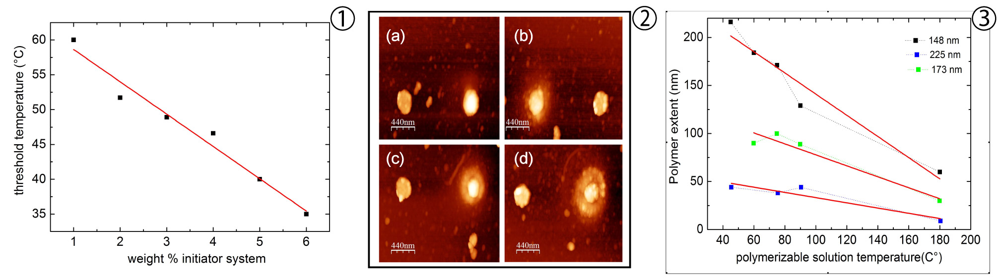
- Fig. 1: Linear variation of the threshold temperature of polymerization as a function of the percentage weight of the initiator system
Fig. 2: AFM images of the nanothermopolymerization in the vicinity of single nanoparticles photo excited at different time from 1 min (a) to 13 min (d)
Fig. 3: Variation of the polymer extent according to the threshold temperature of the various polymerisable solutions in the vicinity of three types of nanoparticles having different resonance Plasmon position.
[1] G. Baffou and R. Quidant, Laser Photon. Rev. (2012) 1-17. [2] J. S. Donner et al., Nano lett 12 (2012) 2107. [3] G. Baffou et al., Acs Nano 6(3) (2012) 2452.
1 Humboldt-Universität zu Berlin, Department of Chemistry, Brook-Taylor-Str. 2, 12489 Berlin
2 BAM Federal Institute for Materials Research and Testing, Richard-Willstätter-Str. 11, 12489 Berlin, Germany
3 Technische Universität Berlin, Central Facility for Electron Microscopy (ZELMI), Straße des 17. Juni 135, 10623 Berlin, Germany
Investigating the interaction of nanomaterials with biomolecules or cells is of great interest, since nanoparticles are omnipresent in the environment and are used for various applications in biotechnology and medicine.
Nanostructures of gold or silver combined with silica shells (SiO2@Au, SiO2@Ag) are attractive systems for many uses as they combine the physical and chemical properties of two different materials. The plasmonic cores can serve as optical sensors, e.g., in analyses using surface-enhanced Raman scattering (SERS). Compared to uncoated nanoparticles, the coating with silica changes the surface properties and also many other characteristics of the nanoparticles in complex biosystems.
Here, we present results of synthesis and characterization of new SiO2@Au and SiO2@Ag nanostructures. Experimental parameters of the silica coating procedure were varied to adapt the particle properties to the requirements of their future application in cells. Plasmonic nanoparticles with silica layers of different thicknesses were synthesized. Prior to in vitro studies, the SiO2-nanocomposites have to be well characterized with respect to layer thickness and stability, e.g., in cell culture medium.
We acknowledge funding by ERC starting grant No. 259432 (MULTIBIOPHOT) and Einstein Foundation Berlin.
1 Institute of Photonic Technology Jena (IPHT), Albert-Einstein-Straße 9, 07745 Jena, Germany 2 Institute of Physical Chemistry and Abbe Center of Photonics, Friedrich-Schiller-University, Helmholtzweg 4, 07743 Jena, Germany
This contribution presents a method which enables very fast and simple coating of small (<10nm) silver nanoparticles (AgNP) and there utilization as SERS substrates. The AgNP were synthesized chemically by reduction of Ag+ in aqueous solution. Afterwards, the AgNP were coated with a hydrophobic alkanethiol by phase transfer into a non-polar solvent. The phase transfer can be done without surfactant which keeps the suspension pure for subsequent use. Furthermore, approaches to concentrate and purify the coated particles are shown. Afterwards, the coated AgNP can form SAMs using a solvent exchange method. This allows the formation of tight AgNP monolayers on various substrates which can be dried for subsequent measurements.
The SAMs were characterized by TEM imaging, optical imaging and spectroscopic measurements. Finally, their usability as SERS substrates is demonstrated by the detection of a hydrophobic model analyte (Sudan III), which could be detected by SERS measurements down to 10-5 M.