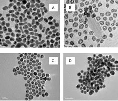
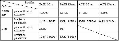
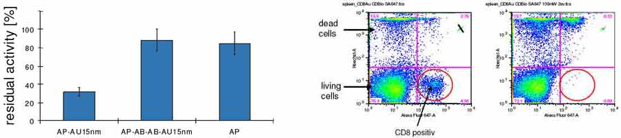
Surface plasmon mediated fluorescence Bill Barnes School of Physics, University of Exeter The emission of light by a molecule is altered when it takes place in proximity to a metal surface. In this talk I will describe the radiative and non-radiative decay pathways that are opened up as a consequence of the presence of the metal, and will discuss how the relative importance of these pathways varies with molecule-surface separation. I will in particular look at the scope for recovering energy lost to non-radiative modes as light. Fabrication and characterization of nanophotonic metal structures A. Csaki, A. Steinbrück, S. Schröter*, T. Glaser*, W. Fritzsche Institute for Physical High Technology Jena, Biotechnical Microsystems Department; *Microoptics Department Metal nanostructures with sub-wavelength dimensions exhibit interesting optical properties. Nanoaperture and nanodot arrays were prepared using electron beam lithography of thin metal layers. AFM imaging was used to determine the lateral dimensions and the topography of the resulting structures. Beside an optical characterization, the work was focused on a possible combination of these structure with metal nanoparticles originating from chemical synthesis. Such nanoparticles were immobilized on nanoaperture arrays, and individual apertures with a single particle could be identified and characterized using both ultrastructural and optical methods. Figure: Subwavenlength nanoapertures fabricated in thin metal films. Left: Optical micrograph of an aperture array. Center: Zoom by optical microscopy and AFM. right: One aperture (center) filled with a 30 nm nanoparticle, three other particles are also visible. AFM image. The Extraordinary Transmission of Metallic Arrays of Subwavelength Apertures for Enhanced IR Absorption Spectra of Complex Molecular Assemblies Shannon Teeters-Kennedy, Kenneth R. Rodriguez, Shaun M. Williams, Alexandra Sudnitsyn, Summit Shah, Frank Hrovat, and James V. Coe The Ohio State University Nanoscale assembly efforts can be facilitated with simple spectroscopic methods to characterize surfaces. Ebbesen's extraordinary transmission of biperiodic metal arrays of subwavelength holes (1) has been moved into the infrared (IR) region by using metal arrays with larger spacings (hole widths of ~3 m and hole-to-hole spacing of ~13 m on a square lattice) in order to get transmission over the traditional range of molecular vibrations. This effect is thought to be mediated by surface plasmons (although some are suggesting a role for evanescent diffracted waves) and this new method for accessing surface plasmons in the mid-IR presents the possibility of working with a much wider range of metals than are typically used in visible surface plasmon studies (such as Ni, W, Pt, Pd, and Cr, as well as Cu, Ag, and Au which are typically used in the visible). Using standard FTIR instrumentation, a method has been developed to record enhanced infrared absorption spectra of metal-supported self-assembled monolayers, phospholipid bilayer coatings, and membrane bound proteins supported within stacks of such systems. Enhancements in absorbance of 100 to 1000-fold have been observed in alkanethiol self-assembled monolayers over literature reports (2). The basic optical physics as well as several potential applications (3) will be discussed. 1 T. W. Ebbesen, H. J. Lezec, H. F. Ghaemi, T. Thio, and P. A. Wolff, Nature (London) 391(6668), 667 (1998). Novel Fluorescent and Raman-Active Noble Metal Quantum Dots Robert M. Dickson School of Chemistry and Biochemistry, Georgia Institute of Technology, Atlanta, GA30332-0400 Highly fluorescent and Raman active, water-soluble, several-atom gold and silver quantum dots have been created in dendritic and peptide matrices and are readily observed on the single molecule level. These quantum dots behave as multi-electron artificial atoms with size-tunable, discrete electronic transitions between states of welldefined angular momenta. Correlation of Au nanocluster size with transition energy is well-fit by the simple relation, Efermi/N1/3, indicating protoplasmonic fluorescence arising from intraband transitions of free electrons (the jellium model). These conduction electron transitions are the low number limit of the plasmon - the collective dipole oscillations occurring when a continuous density of states is reached. Photon antibunching experiments further indicate that single electron transitions instead of collective oscillations are responsible for the size-dependent emission. Providing the missing link between atomic and nanoparticle behavior in noble metals, these highly emissive, water-soluble noble metal quantum dots offer complementary transition energy size scalings at smaller dimensions than do semiconductor quantum dots. The unique, discrete excitation and emission coupled with facile creation in aqueous solution open new opportunities for noble metal quantum dots as biological labels, energy transfer pairs, and other light emitting sources in nanoscale optoelectronics. 3D Mid-Field Microscope: Future Application Of Extraordinary Trans-Mission Through Subwavelength Hole-Arrays M.W. Docter, I. T. Young and Y. Garini Imaging Science and Technology, Delft University of Technology Extra ordinary transmission [1] through a single hole in a metal occurs when this hole is periodically arranged with other holes or grooves. The resulting transmission exceeds unity, it is spectrally selective and most important for our application, there is a small angular diffraction. In our 3D mid-field microscope the transmission through a periodic array will be used to illuminate a biological sample in near-field. Fluorescent molecules will be used as probes, which will allow us to label and image the sample in 3D. The most important advantage of this microscope originates from the low diffraction of light through a relatively thick sample with enhanced transmission. Therefore, the samples 3D interior can be measured, while a near-field microscope is limited to only the 2D surface of the sample. The resolution and depth of field of the mid-field microscope strongly depend on the angular diffraction. We will examine this angular diffraction by conducting both near-field and far-field measurements. Each method has its own advantages and disadvantages. Far-field measurements that we already performed [2], have the advantage that the measurement does not interfere with the plasmon field. Preliminary results are shown in figure 1. Near-field measurements will enable us to achieve higher spatial resolution though it has the disadvantage that the scanning tip itself effects the electric field in the scanning vicinity. The results in both far and near-field will be presented in more detail and their key role in the development of the mid-field microscope will be discussed. Figure 1. Far-field measurements on transmission through a 25x25 hole array, pitch 700 nm, diameter 150 nm. A-C: Koehler illum. (collection of angles); B-D: Collimated illumination (angle = 0°); A-B: 450-550 nm; C-D: 650-750 nm. [1] T.W. Ebbesen et al., Nature 391, 667 (1998) Fluorescence radiation engineering with metallic nanocavities Jörg Enderlein IBI-1, Forschungszentrum Jülich, 52425 Jülich A theoretical study of the fluorescence properties of fluorophores within nanometric metallic nanocavities is presented. The study is based on a semi-classical approach solving exactly Maxwell's equations and using Fermi's golden rule. Several effects are taken into account: Local field enhancement of the fluorescence-exciting electromagnetic field, fast energy transfer from the fluorophore's excited state to metal nanocavity plasmons, re-emission of light from the excited cavity's plasmons, energy losses due to optical absorption and dissipation by the cavity. It is shown the presence of the metallic nanocavity influences all aspects of the fluorescence emission, in particular brightness (effective absorption cross section), lifetime, absorption and emission spectra, and effective quantum yield. The connection between fluorophore emission and changed vacuum field structure is also briefly discussed. DNA-based nanoparticle plasmonics for a highly parallel and integrated molecular nanotechnology Wolfgang Fritzsche Institute for Physical High Technology Jena, Biotechnical Microsystems Department Detection and manipulation of molecules is a key issue in a wide range of research fields, ranging from molecular diagnostics over single molecule techniques to molecular nanotechnology. Bioconjugated metal nanoparticles, especially those complexes made by DNA, represent a novel, effective tool for a highly sensitive and - at the same time - comparatively straightforward detecion of molecules with optical methods. The methods established for preparation but also manipulation and characterization of these bioconjugates can be applied in the development of novel optical nanoparticle-based techniques. The nanoparticles are utilized as nanoantennas, small structures that enable highly localized energy conversion with the ability to either bring thermal energy into nanoscale structures or even to induce local destruction.On the other hand, the described nanoparticle-DNA conjugates provide a platform for highly defined functional units with interesting optical properties such as wave guides or molecular beacons. The synthesis based on self organization enables high parallelization and thereby the potential for widespread future application. However, a synthesis in solution also results in the typical integration problem: How to interface and combine these units with a technical environment, in order to access and use the functionality. Multiple-step biomolecular self-assembly using positioned extended DNA molecules could overcome this problem, if it can be based on parallel techniques. Adapting DNA stretching methods to microstructured substrates allowed for the parallel positioning of individual DNA structures in electrode gaps, and these structures were successfully functionalized using specific binding of nanoparticles. This development will enable a nanophotonic based on methods from microsystem technology and DNA nanotechnology for further developments in ultrasensitive bioanalytics as well as nanooptics. Laser-based sequence-specific DNA processing with sub-wavelength precision using DNA-nanoparticle conjugates A. Csaki, F. Garwe*, A. Steinbrück, A. Weise#, G. Maubach, K. König*, W. Fritzsche Institute for Physical High Technology Jena, Biotechnical Microsystems Department; *JenLab GmbH, Jena; #Institute for Human Genetics and Anthropology, Friedrich-Schiller-University Jena DNA restriction is a basic method in today's molecular biology. Besides application for DNA manipulation, this method is e.g. used in DNA analytics for 'restriction analysis'. Thereby DNA is digested by sequence specific restriction enzymes, and the length distribution of the resulting fragments is detected by gel electrophoresis. Differences in the sequence lead to different restriction patterns. A disadvantage of this standard method is the limitation to a small set of known restriction enzymes with fixed sequences, so that the assay cannot be adapted to any sequence of interest (e.g. SNPs).We designed a scheme for DNA restriction in order to provide access to any desired sequence, based on laser light conversion on sequence-specific positioned metal nanoparticles. Especially gold nanoparticles are known for their interesting optical properties caused by plasmon resonance. The resulting absorption can be used to convert laser light pulses into heat, resulting in nanoparticle destruction. We work on the combination of this principle with DNA-modification of nanoparticles and the sequence-specific binding (hybridization) of these DNA-nanoparticle complexes along DNA molecules. Different mechanisms of light-conversion were studied, and the destructive effect of laser light on the nanoparticles and DNA was demonstrated. Laser irradiation parameters like power density or number of pulses were varied in order to find optimal conditions for damages confined to the particles, but to realize otherwise unharmed samples. This could be demonstrated with sequence-specific bound nanoparticles on human metaphase chromosomes using an ultrastructural characterization of the laser effect by AFM imaging the binding region before and after laser irradiation. Further work will address the study of individual DNA molecules and their controlled manipulation using this technique. Figure: AFM images of human metaphse chromosomes with sequence-specific nanoparticle label (arrow) before (left) and after (right) laser irradiation. The zoom images (insets) show clearly the highly localized damaging effect. Funded by BMBF (FKZ 0312013B, NanoCut) in the framework of the "Nanobiotechnology" initiative. A. Csaki, G. Maubach, F. Garwe, A. Steinbrück, K. König, W. Fritzsche: A novel DNA restriction technology based on laser pulse energy conversion on sequence-specific bound metal nanoparticles. Proc. SPIE 2005 Vol. 5699, p. 436-441. Application of Laser irradiated gold nanoparticles for selective protein knock out, cell permeabilisation, and cell killing Gereon Hüttmann, Marco Bever (Institute for Biomedical Optics, University of Luebeck, Germany), Benno Radt (Carl Zeiss AG, Jena), Ramtin Rahmanzadeh, Johannes Geerdes (Research Center Borstel, Germany), Cuiping Yao (Xi'an Jitotong University, Xi'an, China), Elmar Endl (Institute of Molecular Medicine and Experimental Immunology,University Bonn, Germany) The strong absorption of gold nanoparticles in the visible spectral range allows the localized generation of heat in a volume of only a few tens of a nanometer. Irradiation with a pulsed lasers can easily heat up the particles above the melting temperature of gold. Due to their small volume cooling by heat diffusion is very efficient. Therefore temperatures above 1000°C can be confined to the surroundings of the nanoparticles with heating times in the sub-nanosecond range. Pulsed irradiation of gold nanoparticles is therefore expected to cause very localized chemical, thermal or mechanical modifications to cells and biomolecules. We demonstrate the selective destruction of proteins, the permeabilization of the cell membrane and selective killing of cells by laser-irradiated gold nanoparticles.As a model system for investigating the effect of laser-irradiated gold nanoparticles on proteins, conjugates were made from the different enzymes (alkaline phosphatase or chymotrypsin) with nanoparticles of different size (6 nm, 15 nm and 30 nm diameter). We were able to show, that an inactivation of the enzymes is possible with very high spatial confinement (Fig. 1).Permeabilization was studied systematically in different cells lines. antibodies against membrane proteins were conjugated to gold nanoparticles. The conjugates than were bound to the cell membrane and subsequently irradiated by nanosecond and picosecond laser pulses. Transient permeabilization was observed for 10 kDa Dextran. Efficacies of more than 60% were attained under optimal conditions with only 27% cell death (Table 1). These results show, that a selective transfer of macromolecules across the plasma membrane can be induced by laser irradiated gold nanoparticles. Table 1: The best efficacies for the transfer of 10 kDa FITC-Dextran into Karpas 299 and L428 cells with the corresponding irradiation parameter. When irradiated with higher pulse energies, cells to which the gold particles were bound, were effectively killed. In mixed cell cultures of different lymphoma cells, the targeted cells were killed with over 95% efficacy without significantly affecting the non-targeted cells. an elimination of CD8 positive cells was also demonstrated in spleen cells which were freshly harvested form mice (Fig. 2). This experiment shows, that the selective elimination of cell is not only possible in cell culture. Fig. 1 (left): Enzyme activity of alkaline phosphatase (AP) coupled directly to 15 nm gold nanoparticles (AP-AU15nm), coupled via two antibodies (AP-AB-AB-AU15nm), and without gold particles after irradiation with picosecond laser pulses. Fig. 2 (right): Selective targeting of CD8 positive T-cells from mouse spleen by laser-irradiated gold nanoparticles. Identification of the CD8 positive cells und measurement of cell viability was performed by flow-cytometry Left: Dot diagram of a mixture of spleen cells before irradiation, which shows a certain fraction of CD8 positive cells. Right: Dot diagram after irradiation. CD8 positive cells were killed completely. In combination with selectively binding antibodies, laser-irradiated Gold nanoparticles allow a precise and effective destruction of biomolecules and cells, which can be used for a new kind nanoparticle mediated cell surgery (NPCS). Possible application are protein knock-out, cell purging or a selective depletion of cells from cell or tissue culture. Surface-enhanced Resonance Raman Scattering (SERRS) and fluorescence near metal nanoparticles Peter Johansson1, Hongxing Xu2, Mikael Käll3 1Dep. of Nat. Sciences, Univ. Örebro, Sweden; 2Division of Solid State Physics, Lund Univ., Sweden; 3Dep. of Applied Physics, Chalmers Univ. of Techn., Göteborg, Sweden We present a general model study of SERRS and surface-enhanced fluorescence, focusing on the interplay between electromagnetic (EM) enhancement effects and the molecular dynamics, as treated by a density matrix calculation [1,2]. The model molecule has two electronic levels and one vibrational mode, is affected by radiative and non-radiative damping mechanisms, and a Frank-Condon mechanism yields electron-vibration coupling. Fig. 1. Calculated spectra for the model R6G molecule at three different excitation energies and an incident intensity of 0.13mW/mm2. The molecule is situated symmetrically between two Ag spheres (R = 40 nm) at a distance of 5 Å from the surface. The sharp peaks at the excitation energies is the molecular Rayleigh scattering while peaks at lower energies are 1:st and 2:nd order Stokes Raman scattering. The broad continuum is the fluorescence contribution, which is not completely quenched because of the high field enhancement between the Ag particles. [1] H.X. Xu et al., PRL 93, 243002 (2004) Striped metal nanowires: optical properties and applications Christine D. Keating Department of Chemistry, Penn State University, University Park, PA 16802, USA. Nanowires having stripes of different metals can be prepared by sequential electrochemical deposition within the pores of alumina membranes. After release from the membrane templates, nanowires can be coated with organic or inorganic films. We have prepared linear chains of nanoparticles with controlled interparticle spacing by selective etching of SiO2-coated Au/Ni or Ag/Ni wires. The optical properties of such nanoparticle chains are dependent upon the size, shape, composition, and spacing of the remaining nanoparticles. These structures, which are prepared in batches of millions to billions via wet chemical methods, are potentially interesting as plasmonic waveguides. When nanowires are not etched, the different metals can be used as an optical 'barcode' to identify nanowires from different batches. When functionalized with antibodies or oligonucleotides, these encoded nanowires can serve as individual biosensors. The metal striping pattern can be identified via the differential reflectivity of adjacent stripes using conventional light microscopy. Thus, different sensor nanowires can be mixed together to perform multiple, simultaneous bioassays. Reflectivity-based readout of particle patterns does not interfere with the use of fluorescence for detection of analytes bound to particles by affinity capture. Because they enable testing for multiple targets at once, without specialized instrumentation, these barcoded nanowires have potential for clinical pathogen diagnostics. We have explored the interactions between fluorescent tags used in bioanalysis and the metal surface of the nanowires as a function of wavelength, metal, dye molecule, and metal-dye separation. Optical Probes for Biological applications Based on Surface Enhanced Raman Scattering on Gold Nanoparticles Janina Kneipp, Harald Kneipp, William L. Rice, Katrin Kneipp Harvard University, Medical School, Wellman Center for Photomedicine, Boston, MA Current biochemical and cell biology research generates a strong need for improving optical probes regarding sensitivity, specificity, molecular structural information content, and spatial localization. In particular, probes which can deliver chemical structure information from a biological environment would be of enormous advantage.Metal nanostructures open exciting new ways to create efficient optical probes, based on the strongly enhanced spectroscopic signals that can occur in their local optical fields. One of the most impressive effects associated with local optical fields is surface enhanced Raman scattering (SERS). Total SERS enhancement factors can reach 14 orders of magnitude, which brings non resonant surface enhanced Raman signals to a level comparably to or even better than fluorescence. Unlike fluorescence, which produces relatively broad bands, Raman scattering as a vibrational, i.e., structure-specific method yields a unique spectrum composed of several narrow spectral lines, resulting in well distinguishable spectra even for similar molecules.Here we propose a probe based on the SERS signal of the dye indocyanine green (ICG) on gold nanoparticles and demonstrate the application of this biocompatible probe in living cells. The probe can be detected by the characteristic ICG SERS signature. as our data indicate, the SERS spectrum of ICG consists of more than ten characteristic bands distributed over a broad frequency range. For imaging of the label, this offers the advantage that spectral correlation methods can be used to enhance the contrast between the label and the cellular background. at the same time, an ICG gold nanoprobe is capable of delivering spatially localized chemical information from the cells by employing SERS in the local optical fields of the gold nanoparticles. although the Raman shifts, relative peak intensities, and line widths with SERS may slightly differ from those in normal Raman spectra due to the interaction of the molecule with the metal and due to large field gradients, many bands show similarities with spectra collected from cell constituents in normal Raman measurements. However, compared to normal non resonant Raman experiments, the large effective scattering cross section in SERS allows application of very low laser powers (< 2mW at 830 nm focused to ~1 μm) and very short data acquisition times of 1 second and less, both major prerequisites for reliable in vivo studies.The potential of this kind of SERS probes for controlled targeted vibrational characterization of biological systems will be discussed. Dyes and optical antenna M. Kreiter, K. Vasilev, F.D. Stefani, J. Shumaker-Parry, H. Rochholz, M. Stemmler, F. Gaul Max Planck Institut für Polymerforschung, Ackermannweg 10, 55128 Mainz, Germany The interaction of chromophores with plasmonic nanostructures may lead to an increase in dye performance in terms of absorption cross-section, photostability, and quantum efficiency. For a quantitative understanding of these effects, well-defined nanoscopic geometries are required.First, experiments in an ultraflat planar multilayer structure with chromophores being separated from a thin gold film by a dielectric spacer are discussed. It is shown that, due to surface-plasmon mediated excitation and emission, single-molecule imaging through the film is possible and even more efficient in the presence of gold compared to the pure dielectric case (See Fig. 1). a single-molecule study reveals an influence of the metal not only on singlet decay but although on intersystem crossing. The influence of the metal on intensity and photostability is investigated in ensemble studies.Novel, crescent-shaped gold nanostructures prepared by colloidal lithography are presented (See Fig. 2) that support several strong plasmonic resonances in the near infrared which may have at least partly magnetic character and show strong potential for biosensing. Figure1: Calculated (left) and measured (right) pattern of a single fluorophore as seen by a scanning confocal microscope through a thin gold film. Influencing a single fluorescent molecule with a single metallic nanoparticle S. Kühn, U. Hakanson, L. Rogobete and V. Sandoghdar Laboratory of Physical Chemistry, Swiss Federal Institute of Technology (ETH), CH-8093 Zürich, Switzerland We investigate the influence of a single metallic nanoparticle on the emission properties of a single fluorophore. We use a shear-force based scanning near-field optical microscope (SNOM) to position a single gold nanoparticle in front of an ultrathin crystalline film, containing orientend molecules. By measuring the emission intensity and fluorescence lifetime, we study the modification of the optical properties of a single molecule due to its interaction with the gold nanoparticle.The effect of surface enhancement of emission has motivated a vast number of experimental and theoretical efforts by numerous physicists, spectroscopists and analytical chemists for more than thirty years. a few groups have reported Raman scattering enhancement factors of up to 1015 at the single molecule level while there is still much debate about the physical origin of this effect and the conditions for its reproducibility. Traditional experiments suffer from a double ensemble averaging taking place over the optical response of an extended nonuniform surface, on the one hand, and over the locations and orientations of analyte moleceules on the other hand. Because it is expected that the very local details of the enhancing substrate geometry and material as well as its separation and orientation with respect to the analyte play signifcant roles, we have set out to perform experiments that allow us to control these parameters with precision and at will. The starting point in our work is the attachment of a gold nanoparticle to the end of a glass tip [1]. We then record the plasmon resonance of this probe and approach it to a sample containing oriented terrylene molecules [2]. at each scan pixel we record the fluorescence intensity (Fig. 1) as well as the fluorescence lifetime or spectrum for different excitation wavelengths and polarizations. We observe profound changes of the fluorescence properties of the molecule. By choosing different guest molecules we are able to switch the molecular orientation discretely from perpendicular to parallel to the sample surface. The results are found to be in good agreement with the predictions of theoretical models. In addition, tip-induced changes of the photophysical behavior like intersystemcrossing and photobleaching are observed.Single walled carbon nanotubes are utilized to evaluate the capability of achieving surface enhanced Raman scattering (SERS) with our tips. We will discuss the implications of our investigations for the role of field enhancement in SERS. Figure 1: a) Schematics of our experimental arrangement, b) Flourescence intensity and excited state lifetime as a function of its separation from the gold nanoparticle [1] T. Kalkbrenner, M. Ramstein, J. Mlynek and V. Sandoghdar, J. Microscopy 202, 72 (2001). Metallic Nanostructures for High Sensitivity Fluorescence Detection Joseph R. Lakowicz Center for Fluorescence Spectroscopy at the University of Maryland School of Medicine, 725 West Lombard Street, Baltimore, MD 21201 Recently we demonstrated the possibility of fluorescence enhancements near metallic nanostructures, which result from radiating plasmons. The magnitude of enhancement depends on the size and shape of metallic particles. Theoretical predictions and experimental results demonstrate strong fluorescence signal enhancements of the emission of fluorophores positioned close to the metallic particles and structured nanosurfaces. This phenomenon has been named Radiative Decay Engineering (RDE). The study of single molecule emission on silver island film demonstrates the extraordinary enhancements potential. RDE provides many possible applications in biological assays and DNA arrays by enhancing the dynamic range of sensitivities. Examples of model immunoassays and DNA hybridizations will be presented. We also present a related phenomenon called Surface Plasmon-Coupled Emission (SPCE). SPCE gives the possibility of directional rather than isotropic emission and offers an exceptional background rejection. SPCE has been studied on various metallic semitransparent mirrors, and provides efficient collection of a large fraction of the total emission. Figure 1. Silver island film enhances the emission of YOYO-labeled DNA 15-fold. Optical Properties Of Gold Nanorods: Orientation Effects and Mechanical Oscillations Paul Mulvaney Stiftung Caesar, Ludwig Erhard Allee 2, 53175 Bonn, Germany & School of Chemistry, The University of Melbourne, Victoria 3010, Australia In this talk, we examine the optical properties of gold nanorods, some of their unusual physical properties and their behaviour when excited by short laser pulses. Gold nanorods may be grown in a predictable and tunable way in a single step using wet chemical procedures [1]. Such small metal rods have tunable optical absorption bands and may have potential uses as optical filters. It is important to know what determines the linewidth of the absorption bands and also how fast energy and by what mechanisms optical energy can be dissipated [2]. We show that the rods can be oriented in polymer films and this leads to the creation of simple polarizers. However the photochemical melting of the rods may limit their applications in optical devices [3].In the second part of the talk, we show that at low laser fluxes, irradiation excites vibrational modes within the rods. The breathing mode dominates the mechanical deformation of the rod and this is confirmed by finite element analysis. However, because the frequency of the fundamental extensional mode is much lower than that of the breathing mode, the extensional mode will dominate the transient optical response for a real experiment, that is, for a finite-time heating/expansion process [4,5]. The results of this model are compared to data from transient absorption experiments performed on gold nanorods with average aspect ratios (length / width) between 2 and 6, and lengths between 30 and 110 nm. The transient absorption traces show pronounced modulations with periods between 40 and 120 ps, which are only observed when the probe laser is tuned to the longitudinal plasmon band of the sample. The best fit suggests the Young's modulus is significantly less than that of the bulk material. [1] Perez-Juste, J.; Carnie, S.; Chan, D.Y.C.; Liz-Marzan, L.M.; Mulvaney, P.; Adv. Func. Mat. 14, 571-79 (2004). Tip-enhanced single molecule Raman spectroscopy Markus B. Raschke Max-Born-Insitut, Berlin The optical local-field enhancement at a sharp metallic tip in combination with resonance Raman spectroscopy provide an optical scanning probe method with all-optical ultrahigh spatial resolution down to the several nanometer range and the chemical sensitivity provided by vibrational spectroscopy. Here, illuminating the apex of a au wire tip in close proximity to the sample the vibrational resonance Raman response for submonolayer molecular coverages on planar gold surfaces has been studied.The strong near-field coupling between au tip and sample results in a high degree of field-confinement within a few nm of tip-sample distance and is responsible for a high Raman intensity enhancement. Spectral Raman line narrowing compared to the ensemble average and spectral diffusion is observed for Rhodamine 6G and Malachite green molecules. These temporal fluctuations of spectral position and relative peak intensities as well as transient line splitting in time series of sequentially recorded spectra are indicative of probing only a single emitter in terms of single molecules or small clusters. These results indicate that with further improvement in probe tips and high illumination/detection efficiency single molecule Raman microscopy and spectroscopy is achievable in scattering-type near-field microscopy on arbitrary surfaces. Interactions and morphology of DNA-linked gold nanoparticle networks Ulrike Rehn, Paul Miclea*, Ralf B. Wehrspohn*, and Ulrich Gösele Max Planck Institute of Microstructure Physics, Halle; *Department of Physics, University of Paderborn Complex three-dimensional particle networks can be created via DNA linking of nanoparticles. We analyzed in detail the interactions of double-stranded DNA with gold nanoparticles and the resulting morphologies via UV/Vis-spectroscopy and transmission electron microscopy (TEM). UV/Vis spectroscopy is a powerful method to study DNa-linked gold nanoparticles. The predominant advantage of this method is the concomitant, non-destructive analysis of the DNA status and the particle characteristics. DNA exhibits an absorption maximum at 260 nm, whereas the particle plasmon resonance is in the spectral range between 500 to 600 nm depending on particle size. Therefore, melting of DNA in such hybrid materials or interactions between DNA and particle surface can be analyzed. Beside that, UV/Vis-spectra provide information on the size of generated aggregates. These data have been cross-checked by theoretical calculations using Mie theory and the network have been visualized by TEM analysis.In particular, we studied the interactions and the morphology of particle networks using double-stranded DNA which allows distinguishing between hybridization- or salt effects and bonding of the DNA to the particle. We evidenced that DNA serves as spacer molecule when it is bonded via thiol-groups to the particles. We were able to create particle networks with difference sizes of gold particles (20, 50 and 80nm). It turns out that the particle diameter determines the overall aggregate size: the bigger the particles, the smaller the aggregates. The detailed understanding of the mechanisms of DNA/nanoparticle coupling is crucial for the development of advanced technologies using DNA-hybrid materials as building blocks. Application of different SERS substrates for the investigation of biological samples P. Rösch, R. Geßner,* M. Harz, U. Neugebauer, M. Schmitt, W. Kiefer* and J. Popp Institut für Physikalische Chemie, Friedrich-Schiller-Universität Jena, Helmholtzweg 4, 07743 Jena, Germany; * Institut für Physikalische Chemie, Universität Würzburg, Am Hubland, 97074 Würzburg, Germany For the investigation of biological samples besides microscopic images, it is also important to yield information about the chemical composition of the investigated tissue. In most cases the chemical substances of interest are extracted and analyzed with gas chromatography or HPLC. With these techniques no information about the spatial distribution of the substances within the samples can be obtained. Micro-Raman spectroscopy offers chemical information with a high spatial resolution up to the micrometer range. In addition Raman spectroscopic measurements are not getting perturbed by water which is almost present in all biological samples. However, due to the low concentration of the investigated substances inside the cells the Raman signals are often of low intensity and additionally may be masked by fluorescence. These problems can be avoided by applying the surface enhanced Raman spectroscopy (SERS). The enhancement effect is due to two possible mechanisms: a chemical enhancement due to charge-transfer-effects of the adsoded molecule and the substrate and an electromagnetic enhancement due to plasmon resonance effects. SERS spectroscopy has the advantage to both enhance certain modes of the molecules of interest and at the same time quench the fluorescence. The most commonly used SERS substrate are silver ore gold colloids which are easy to prepare. However, biological samples may be modified or even destroyed due to electrochemical reactions with the colloidal metal particles. Therefore, a minimal exposure of the biological samples to the SERS substrate would be favorable. One possibility to design a SERS substrate with minimal invasive characteristics is to localize the colloid only on the measuring position. This can be done e.g. with an etched glass fiber tip. For excitation the laser is coupled in the glass fiber which yields a spatial resolution in the sub micrometer range. Using a silver island film or localized silver colloids on the fiber tip very low laser power can be used due to the enhancement of the SERS substrate. Therefore the biological sample will not be modified according to high laser powers or high amount of colloids in the tissue. In this work we describe the application of such an SERS probe to investigate biological samples. Acknowledgment: The funding of the research project FKZ 13N8369 within the framework 'Biophotonik' from the Federal Ministry of Education and Research, Germany (BMBF) is gratefully acknowledged. [1] R. Geßner, P. Rösch, R. Petry, M. Schmitt, M. A. Strehle, W. Kiefer and J. Popp, "The application of a SERS-fiber probe for the investigation of sensitive biological samples", Analyst 2004, 129, 1193-1199. Enhancement of Fluorescence-based Biosensors based on Localized Surface Plasmon Resonance (LSPR) T. Ruckstuhl, C. McDonagh, O. Stranik, R. Nooney, and B.D. MacCraith Optical Sensors Laboratory, Biomedical Diagnostics Group, National Centre for Sensor Research, Dublin City University, Glasnevin, Dublin 9, Ireland We report on the so-called plasmonic enhancement effect, whereby the presence of metallic nanoparticles or nanostructures in the vicinity of a fluorophore can dramatically alter the fluorescence emission and absorption properties of the fluorophore. The effect, which is associated with the localized surface plasmon resonance (LSPR) of the metallic nanoparticle/nanostructure, depends on parameters such as metal type, particle size and shape and multilayer structure, fluorophore type and fluorophore-particle separation. It has been established that carefully controlled exploitation of the interaction between the LSPR and nearby fluorophores can result in significant enhancement in the performance of fluorescence-based sensors such as biochip platforms.This work is focused on establishing rational design rules for such applications. With this in mind, we have embarked upon a systematic study of the core phenomena underlying LSPR-enhanced fluorescence. In particular, we have developed a range of model systems which enable us to examine separately the impact of the LSPR on enhancement of excitation and emission. Moreover, our investigation has focused on the critical issue of fluorophore-nanoparticle separation in order to identify the optimal distance or 'sweet-spot' of the phenomenon. a particular feature of this work is the development of preparation methods to enable production of metal nanoparticles and nanostructiures, which facilitate reproducible tunability of the LSPR at the optimal separation. With a view to the eventual implementation of this enhancement process in important applications such as diagnostic biochips, compatibility of the nanoparticle preparation and deposition methods with mass production is an important feature. Metal nanoparticles as molecular rulers and orientation sensor for single molecule biophysics Carsten Sönnichsen(*), Björn Reinhard, Jan Liphardt, Paul Alivisatos University of California, Berkeley and Lawrence Berkeley National Laboratory, Berkeley, CA 94720, USA; (*) present address: Physikalische Chemie, Universität Mainz, Welderweg 11, 55099 Mainz Molecular rulers based on Förster Resonance Energy Transfer (FRET) that report conformational changes and intramolecular distances of single biomolecules have helped to understand important biological processes. However, these rulers suffer from low and fluctuating signal intensities from single dyes and limited observation time due to photobleaching. The plasmon resonance in noble metal particles has been suggested as an alternative probe to overcome the limitations of organic fluorophores and the coupling of plasmons in nearby particles has been exploited to detect particle aggregation by a distinct color change in bulk experiments. Here we demonstrate that plasmon coupling can be used to monitor distances between single pairs of gold and silver nanoparticles. We use this effect to follow the directed assembly of gold and silver nanoparticle dimers in real time and to study the time dynamics of single DNA hybridization events. These plasmon rulers allowed us to continuously monitor separations of up to 70 nm for more than 3000 seconds. Single molecule in vitro studies of biological processes previously inaccessible with fluorescence based molecular rulers are now possible with unprecedented time and distance range.Orientation sensors are a growing field of interest in single molecule studies. We present an alternative to the conventionally employed polarization imaging of anisotropic fluorescent probes. By monitoring the polarized light scattering from individual gold nanorods in a darkfield microscope, we are able to determine their orientation as a function of time. Large light scattering efficiency occurs near their plasmon frequency along the long axis of these nanorods. Gold nanorods of 10nm x 50nm are easily produced in high quality in aqueous environment using recently developed soft template methods. We demonstrate time resolution of ms and observation times of hours by observing the two-dimensional rotational diffusion of surface immobilized gold rods. By carefully adjusting pH and ionic strength of the liquid environment, gold nanorods precipitate out of an aqueous suspension and stick to a glass surface but retain their ability to rotate. The observed orientational diffusion shows a fast component of about 60ms. In addition we observe 'sticky times' of seconds, where no rotational diffusion occurs, providing evidence of a shallow trap potential. The large signal to noise ratio, chemical and photochemical stability, fast time response and small size of these gold nanorods make them an ideal probe for orientation sensing in many applications in material science and molecular biology, including the study of rotations in molecular motors. The demonstrated time resolution of 3ms and photostability over hours already surpasses comparable fluorescent probes by orders of magnitude. (1) (1) Sönnichsen, C., Alivisatos, A.P. Nano Letters 2005, 5, 301. Preparation and optical characterization of core-shell bi-metal nanoparticles A. Steinbrück, A. Csaki, C.-C. Neacsu*, M. Raschke*, W. Fritzsche Institute for Physical High Technology Jena, Biotechnical Microsystems Department; *Max-Born-Institute Berlin Chemical approaches allow for the synthesis of highly defined metal heterostructures, such as core-shell nanospheres. Because the material of metal nanoparticles determines the plasmon resonance-induced absorption band, the control of particle composition results in control of the absorption band. Metal deposition on gold or silver nanoparticles yielded core-shell particles with modified optical properties. UV-VIS spectroscopy on solution-grown and immobilized particles was conducted as ensemble measurements, complemented by single particle spectroscopy of selected structures. Increasing layers of a second metal lead to a shift in the absorption band, and shell diameter in the scale of the original particle diameter lead to a predominant influence of the core material. The extent of shell growth could be controlled by reaction time or the concentration of either the metal salt or the reducing agent. Besides the optical characterization, the utilization of AFM, SEM and TEM yielded important information about the ultrastructure of the nanoparticle complexes. Plasmonic Nanophotonics For Ultrahigh Density Nano-Storage Din Ping Tsai Center of Nanostorage Research and Department of Physics, National Taiwan University, Taipei 10617, Taiwan The optical response of the local plasmonic structures of the super-resolution near-field optical structures is the key of the near-field optical recording. The developments of the super-resolution near-field optical structures are closely related to the basic principle of plasmonic nanophotonics, and the connections of the near-field and far-field optical interactions of super-resolution near-field plasmonic structures. We use near-field scanning optical microscopy, Z-scan experiments and optical pump-probe system to explore the optical interactions of various super-resolution near-field plasmonic structures. Many interesting local interactions were found. Measurements of transmission and reflectance indicate complicate transition process of the optical interactions of the super-resolution near-field plasmonic structures. Different local optical responses are observed and analyzed. Possible models of these interactions will be proposed and explained. Figures 1(a) and 1(b) are the normal and nano recording marks on GeSbTe phase-change recording thin film of a DVD disk, respectively. Molecular Plasmonics: Origins, Fundamentals, and Sensor Applications Richard P. Van Duyne Department of Chemistry, Northwestern University, Evanston, USA During the last few years, there has been an explosion of interest and activity in the field of nanophotonics. The goal is to control, manipulate, and amplify light on the nanometer length scale. Two approaches have emerged to accomplish this goal. First, photonic crystals have been fabricated which possess a periodic dielectric contrast on the nanometer scale. Second, the properties of the collective electronic excitations in noble metal films or nanoparticles, known colloquially as surface plasmons, have been explored to achieve highly miniaturized and sensitive photonic devices. Recently, the term plasmonics has been coined to describe this rapidly emerging field. Molecular plasmonics deals with understanding the interactions between adsorbed molecules and plasmonic nanostructures. It is anticipated that molecular plasmonics will have a significant impact on many applications, including localized surface plasmon resonance (LSPR) spectroscopy for chemical and biological sensing, sub-wavelength optical microscopy, surface-enhanced Raman spectroscopy (SERS), and nanolithography.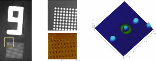

2 K.R. Rodriguez, S. Shah, S.M. Williams, S. Teeters-Kennedy, and J.V. Coe, J. Chem. Phys. 121(18), 8671-8675 (2004).
3 J.V. Coe, S.M. Williams, S.M. Teeters-Kennedy, K.R. Rodriguez, and S. Shah, Nanotechnology, 15 S495-S503 (2004).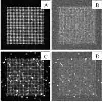
[2] M.W. Docter et al., SPIE Proceedings vol. 5703 (2005, in press)
The model is used to address the case of single-molecule SERRS from fluorescent molecules, specifically the dye molecule Rhodamine 6G (R6G) [3,4]. Realistic molecular parameters are chosen so as to simulate the absorption spectrum of R6G and this model molecule is then placed between two silver spheres, a configuration that provide the most reasonable explanation to the single molecule SERRS effect [5]. The EM enhancement and plasmon modes are found through extended Mie theory. The theory presented here provides an opportunity to calculate actual spectra and absolute cross-sections as a function of incident laser wavelength and intensity, see Fig. 1. Brus and co-workers estimated the maximum integrated spectral cross-section for single Rhodamine 6G molecules on Ag-particle aggregates to 10-14 - 10-13 cm2 [4], which is in excellent agreement with the values obtained from integrating the differential cross-sections in Fig. 1. In the recent SERS literature, e.g ref. [3,4], it is often stated that a surface enhancement of 1014 - 1015 is needed to explain single molecule SERRS. However, we note that the quantitative agreement between experiment and theory found here is obtained through an electromagnetic Raman enhancement of the order 1010 in combination with an ordinary resonance Raman effect for the free molecule. We also note that a broad fluorescence background remains under the Raman peaks even for the shortest metal-molecule distances. It is seems likely that this background gives a major contribution to the "SERS continuum" often reported in the literature, for example in ref. [3,4].
[2] P. Johansson, H.X. Xu, and M. Käll, Phys. Rev. B to appear
[3] S. Nie and S.R. Emory, Science 275, 1102 (1997);
[4] A.M. Michaels et al. , JACS 121, 9932 (1999);
[5] H.X. Xu et al., PRL 83, 4357 (1999).
Figure 2: Gold nanocrescents of varying size and shape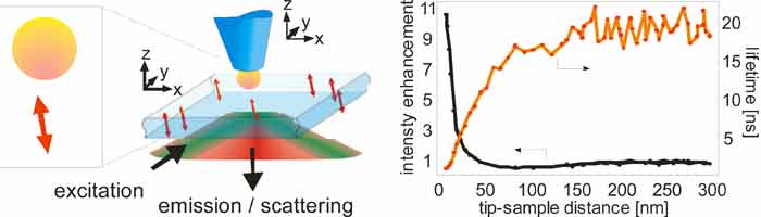
[2] R. J. Pfab, J. Zimmermann, C. Hettich, I. Gerhardt, A. Renn and V. Sandoghdar, Chem. Phys. Lett.387, 490 (2004).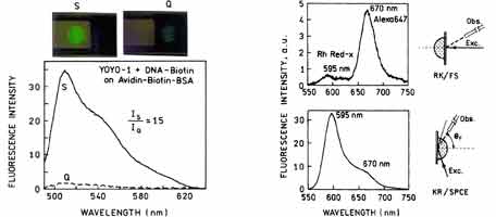
Figure 2. Background suppression in SPCE immunoassay. The molecule of interest is Rh Red-labeled antibody.
[2] Sönnichsen, C; Franzl, T.; Wilk, T.; von Plessen, G.; Feldmann, J.;Wilson, O.; Mulvaney, P.; Phys. Rev. Lett. 77402 (2002).
[3] Wilson, O.; Wilson, G.; Mulvaney, P.; Adv. Mat.14, 1000 (2001).
[4] Hu, M.; Wang, X.; Hartland, G.V.; Mulvaney, P.; Perez-Juste, J.; Sader, J.E.; J. Am. Chem. Soc. 125, 14925-33 (2003).
[5] Perez-Juste, J.; Pastoriza-Santos, I.; Liz-Marzan, L.M.; Mulvaney, P.; Coord Chem Rev (in press 2005).
Plasmonics is a materials driven subject. The unifying theme in this lecture will be the fabrication of size and shape-tunable, silver and gold nanoparticles using nanosphere lithography (NSL) and chemical synthetic methods. Size and shape tunability leads to an exquisite degree of control over the magnitude and spatial extent of the surface electromagnetic fields that surround optically excited nanoparticles. In turn, this has enabled fundamental new insights into the electromagnetic (EM) field enhancement mechanism underlying both LSPR and SER spectroscopy.
This lecture will focus on three topics: (1) the fundamentals of the LSPR; (2) biosensos based on LSPR spectroscopy; and (3) biosensors based on SERS. In the LSPR spectroscopy area we will discuss: (1) the development of nanoscale optical biosensors for the study fundamental biological recognition events and the clinical diagnosis of Alzheimer‘s Disease; (2) the use of chronocoulometry, a quantitative electrochemical method, to „fine tune“ the LSPR; and (3) the release of NSL nanoparticles from the substrate for use as solution phase single nanoparticle sensors. In the area of SERS, we have now mapped out the relationship between the LSPR spectrum of Ag nanoparticles and the wavelength-scanned SER excitation spectra (WS SERES). Application of the WS SERES results to the optimization of SERS-based sensors for glucose monitoring and anthrax detection will be presented.
These results represent important steps in the realization of nanoscale LSPR and SERS sensors for applications in medical diagnostics, biomedical research, environmental science, and homeland defense.